Surgical Pathology:- Part 1- Histopathology, Biopsies
Surgical Pathology
What sample is needed for Surgical Pathology?
- Surgical pathology samples can be obtained from any part of the body, e.g.
- Palpable lumps like:
- breast
- lymph nodes.
- Soft tissue masses.
- Thyroid.
- Palpable lumps like:
- From any visceral organs like:
- Stomach.
- Intestinal Tract.
- Liver.
- Lungs.
- Pleura.
- Abdominal cavity.
- Cervix.
- Endometrial.
- Chorionic villus.
- Kidney.
- Prostate (transrectal biopsy).
- Head and neck.
- Brain.
- Superficial tissues like :
- Oral cavity.
- Skin.
- Eyes.
- Nose and ear.
What are the indications for Surgical Pathology?
- The main purpose of surgical pathology is to diagnose the diseases:
- Inflammatory lesion.
- Neoplastic.
- Benign.
- Malignant.
- A biopsy is needed for the differential diagnosis of the lesion.
- A surgical biopsy is needed to see the resection border for tumor infiltration.
- A biopsy may be done in the follow-up of the tumors.
- Surgical pathology may be needed in the recurrence of the tumor.
- Surgical pathology helps in the staging and grading of malignancies.
- These surgical biopsies are:
- Diagnostic.
- Therapeutic.
What are the types of surgical biopsies?
- Incisional:
- This biopsy is when a piece of the tumor is taken for diagnosis.
- An excisional:
- This biopsy removes the whole tumor and is submitted for surgical pathology reports for diagnosis and resected margins.
- Punch biopsy:
- When the cervix is biopsied.
- A needle biopsy:
- It may be with a wide-bore needle for bone marrow sampling.
- Fine needle when the bore of the needle is 23 to 25 gauge.
- Curettage biopsy:
- When the endometrium biopsy is taken.
What are the surgical pathology procedures?
- Paraffin section (paraffin-embedded method).
- A frozen section (the tissue is frozen and sectioned).
- This is a rapid method and is available during surgical operations.
What are the complications of the biopsy?
- There are chances of bleeding in the case of vascular lesions and the liver.
- There is the possibility of the tumor’s transplantation, which may be seen in the scar area.
- There is a chance of the dissemination of malignant cells.
- There may be perforation during the endoscopic biopsies.
- There is a chance of fistula formation in the pancreas and salivary glands.
How do I send the specimens?
- The biopsy material should be in 10 % formalin, which will prevent the tissue from autolysis. Usually, this is buffered formalin.
- Bouin’s solution.
- Carney’s solution.
- There should be 50% times the biopsy volume.
- Never send the specimen in saline or water.
- If formalin is unavailable, then the simple spirit can be used to avoid autolysis. This is advised only in remote areas and when no other choice exists, particularly in underdeveloped countries.
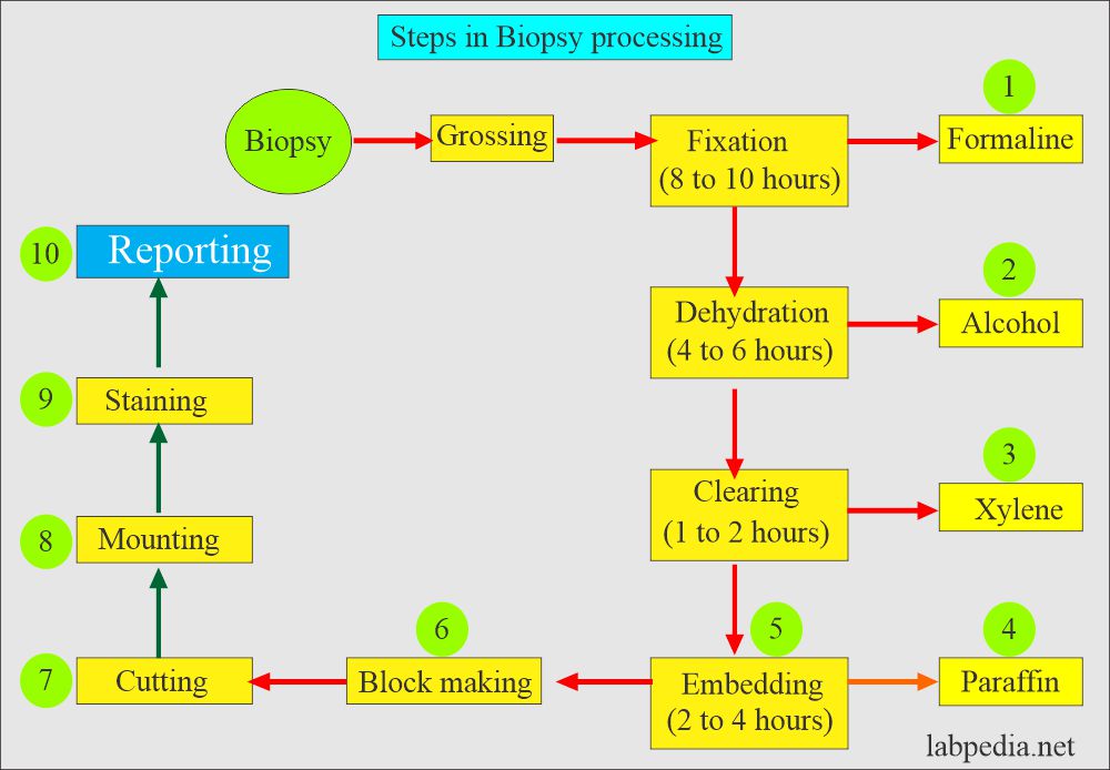
What are the steps in the processing of the Biopsy?
- Fixation:
- The biopsy specimen is kept in formalin for at least 12 to 24 hours.
- This step is very important; if the tissues are not fixed properly, you will not get good slides for interpretation.
- Dehydration:
- The specimen is put into alcohol or acetone in various grades or concentrations.
- This will remove the water and replace it with alcohol. This step takes at least 4 to 6 hours.
- Clearing:
- The specimens are put into xylene, which will replace the alcohol and take its place.
- Now, the tissue is fit for the embedding. This process takes 1 to 2 hours.
- Embedding:
- The specimens are put into melted paraffin (58 °C) and kept for 2 to 4 hours.
- Block making:
- Then, the biopsies are taken from the paraffin, and paraffin blocks are made.
- Cutting:
- The microtome cuts the blocks to a thickness of 4 to 7 microns. The cut sections are then put on the slides.
- Staining:
- Stain the slides with hematoxylin and eosin.
- Mounting:
- The stained slides are mounted with the cover glass.
- Reporting
The surgical pathologist can now read the slides and give his final diagnosis. - It will take at least 24 hours for the biopsy to be done.
Questions and answers:
Question 1: What is the role of formaline?
Question 2: What is the role of xylene?


Would you send me the reference of this article in a pancover way, please.
I’m a medical student and I have a research about biopsy, I need to take some information from this wonderful article.
Please can you explain what you mean by pan cover way?