Lymph node Structure Part 1
Lymph node
Lymph Node are part of the reticuloendothelial system.
Reticuloendothelial System: Reticuloendothelial System is a term used by Aschoff and Kiyono with criteria of:
1. Phagocytic activity.
2. Staining with vital dyes.
3. Juxtaposition to reticulum of connective tissues and lining cells of Blood vessels.
Components of Reticuloendothelial System are :
1. Intravascular:
- · Monocytes
- · Endothelial cells (Kupffer cells)
- · Lymph node sinuses
- · Splenic sinuses
- · Adrenal Capillaries
2. Extravascular:
- · Reticulum cells
- · Histiocytes and macrophages (of Connective tissues)
Lymphoid Tissue
Lymphoid tissue is present throughout the body, calculated that in a man there are about 1300g of lymphoid tissue. Lymph node contains about 100g and bone marrow about 70g.
Lymphoid tissue divided into :
Central components which includes:
- Appendix
- Peyer’s patches
- Thymus
- Tonsils
Peripheral component includes:
- Lymph nodes
- Spleen
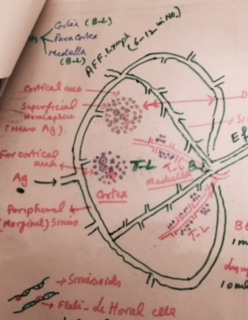
This diagram shows different parts of the Lymph node
Sinusoids are lined by the endothelial cells and in between there are flat Littoral cells.
Flat Littoral Cells has the following properties:
· Can proliferate
· Can Hypertrophy
· Phagocytosis (House cleaning, functions of lymph node)
· May detach and become free phagocytes in lymphoid sinuses and lead to sinus histiocytosis or sinus Catarrh of German authors.
Histology:
Lymph node consists of :
- Medulla.
- Cortex.
- Peripheral sinus.
- Cortical area.
- Deep paracortical area.
- Sinusoides.
- Medullary cards.
- Afferent and efferent vessels
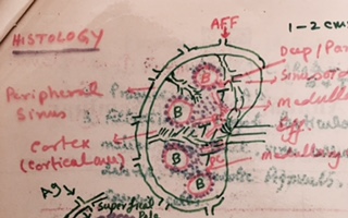
Lymph node showing detail of structure
Primary Follicle when activated, it transform into —-> Secondary Follicle.
Germinal center contains following cells :
- Lymphocytes.
- Lymphoblasts.
- Histiocytes.
- Mitosis.
T- Lymphocytes
T-Lymphocytes converted into à Convoluted T-Cells, this transform into T- Immunoblastic cells, ——->T-Lymphoblast ——> T-Prolymphocytes ——–>T-Lymphocytes.
Functions of lymphocytes:
- Formation of Lymphocytes as B and T lymphocytes which takes part in the immunity.
- Production of Ab takes place by B- lymphocytes.
- Filtration of Lymph removes Particulate matter like Bacteria, Virus, cellular debris, anthracotic pigments and cancer cells.
- Phagocytosis by these lymphocytes.
- They have important role Role in Inflammation and Infection:
Lymph Node are labile. Relatively larger at birth, Modified by stress, adrenal gland , thyroid hormones and immune response.
· Biopsy of Lymph Node used for:
- Bacterial Examination where one can do culture and sensitivity(C/S).
- Cytologic imprint for the better differentiation of the cells.
- Histological Examination can differentiate inflammation, metastatic tumor infiltrate, or primary tumor like lymphomas .
Electron microscopy may be done on biopsy material:
- Immunophenotyping.
- Chromosomal studies.
- Gene Rearrangement Analysis.
- DNA Ploidy studies
Indications for Lymph Node Biopsy:
- To make a diagnoses in case of Persistent enlargement of the lymph nodes
- To make diagnosis in a patient with PUO, weight loss and enlarged lymph nodes.
- To confirm diagnoses already suspected on other grounds.
- To assess in the extent of spread of known malignant diseases.
- To monitor the progress of disease in patient with malignant lymph nodes.
Precaution for lymph node biopsy:
- Inguinal lymph nodes wherever these are enlarged should be avoided. Take biopsy from other group.
- Always take biopsy from the larger lymph node.
- Axillary and cervical lymph nodes are more representative.
- Superficial small lymph node may slow non-specific hyperplasia whereas deeper node in the same group may show diagnostic feature.
- Lymph node with normal picture shows:
- Narrow peripheral sinus.
- Sinusoids are not prominent.
- Macrophages are not prominent.
- There are Small ill-defined germinal centers.
- There are Sparse Lymphocytes in Reticular framework.
- The Size is Small.
Activation of Lymph Node in Acute Disease:
- There is increase in the size of lymph node.
- There is dilatation of the sinusoidal Sinuses.
- The Sinusoids are prominent.
- The Macrophages are increased in number.
- There is increased number of Polys and Necrosis.
- The germinal centers are prominent with increased number of mitosis.
Activation of Lymph Node in Chronic Disease
- There is increase in the size of lymph node.
- There is increase number of plasma cells and eosinophils.
- Also can see above changes seen in acute diseases.