Lymph node, Lymphomas, Non-Hodgkin lymphoma Part 3
Malignant Lymphoma
The lymphomas are monoclonal in origin.
Definition:
Rappaport 1996 Defined that It is neoplastic proliferation of Lymphocyte, Histiocytes and reticulum cells in lymphoid tissue in the body. It is most common in Lymph Node.
Various names:
- These lymphomas are also called Immunoproliferative disorders.
- Britisher calls it Reticulosis, or Reticuloendothelial.
Lymphoma:
This is a Misnomer because it spreads from single Lymph Node to chain of Lymph Node and distant nodes. It also spreads to liver, spleen and Bone marrow where it gives leukemia phase.
Lymphosarcoma:
It is a special variant of NHL (PDL)
Lymphomas are classified into :
1. Non-Hodgkin’s lymphoma
2. Hodgkin’s lymphoma
Confusion for classification is due to:
1. Histogenesis.
2. Degree of differentiation.
3. Failure to differentiate between Lymphocyte and Epithelial cells.
4. Difficult to differentiate malignant or hyperplastic lesion.
Non-Hodgkin’s lymphoma (NHL)
- Non-Hodgkin lymphoma may be localized or generalized.
- 2/3 of Non-Hodgkin lymphoma are primary origin from lymph nodes while 1/3 or extranodal and they may originate from bone marrow, Oropharynx, Gastrointestinal Tract and skin.
- 3% of all malignancies in western countries are non-Hodgkin lymphomas.
Reason for classification of NHL:
- Categorization to know response to treatment.
- Should be easy to be followed by pathologist.
Well known classification of NHL are :
- Rappaport 1996
- Bennett 1974
- Dorfman 1974
- Lukes and Collin 1974
- Lennert
- Mathew 1976
- Working formulation 1982
- Real classification (Revised European-American classification of lymphoid neoplasm) 1994
Non-Hodgkin’s Lymphoma:
- Majority (80-85%) are B-Lymphocyte origin.
- Rest non-Hodgkin’s lymphoma are T-Lymphocyte
- Histiocytic in origin are uncommon.
- Non-Hodgkin’s lymphoma 2/3 are nodal in origin including Hodgkin’s lymphoma while 1/3 are extra-nodal e.g. skin, stomach and brain.
Rappaport Classification:
Given in 1966 and modified in 1978.
Criteria of classification:
- This depends upon;
- Nodular or diffuse
- Cell Morphology, where we look for:
- Size of cells.
- Nuclear shape.
- Nucleoli.
- Cytoplasm.
- Chromatin
- Mitosis.
- Two cell subset (Lymphocytes and Histiocytes).
- Degree of differentiation of cells.
Classification of lymphomas by Rappaport
| Nodular type | Diffuse type | ||
| Poorly differentiated | 24% | well differentiated lymphocytic | 5 % |
| Mixed cell type | 12 % | Poorly differentiated | 16 % |
| Histiocytic | 3 % | Histiocytic | 28 % |
| Mixed cell type | 6 % | ||
| Lymphoblastic | <5 % | ||
| Undifferentiated (Burkitt’s type) | |||
| Undifferentiated (Non-Burkitt’s) |
Good Points of Rappaport:
- Nodular (follicular) or diffuse : This is easy to appreciate and has good prognosis.
- Easy to follow histologically.
Drawbacks:
- Type of cells are not mentioned either B or T-Lymphocyte.
- Transformed activated B-Lymphocytes looks like histiocytes.
- Small B-Lymphocyte end-stage is incorrect.
Rappaport classification is based on cytologic cell division like :
- Small lymphocytes.
- Histiocytes which are 2 to 3 times than the small lymphocytes with 2 to 5 nucleoli and abundant cytoplasm.
- Then are the undifferentiated cells.
Nodular Type Non-Hodgkin’s Lymphoma
- These are 40% of the lymphomas.
- These are the tumor of old age and rear before the age of 20 years.
- Sex ratio is female to male is equal.
- These lymphomas have better prognosis.
- These tumors can transform into diffuse form which take at least 8 years.
- These tumors needs to be differentiated from follicular hyperplastic changes in the lymph node.
- Mitosis are rare.
- The cells are Follicular small cell (FSC) and mixed cells are Small cleaved (SC) and large cleaved cells (LC)>
- Surface markers are :
- Positive surface Ig.
- Positive pan B CD molecule 19 +.
- 75% of the cases bone marrow is involved.
- Extranodal spread is rare.
- Median survival is 7 to 9 years.
Diffuse well differentiated lymphocytic lymphoma
- These are low grade and are 5% of the lymphomas.
- 40% are the transform into chronic lymphocytic leukemia.
- There are small B- lymphocytes with compact nuclei.
- The mitosis are rare.
- These are the tumor of old age.
- There is monoclonal gammopathy.
- Tumor marker shows surface Immunoglobulin Igm and IgD.
- These cells are CD19 + cells.
- Bone marrow is almost always positive.
- These are the low grade lymphomas.
Poorly differentiated Lymphoma (PDL)
- These can be :
- Nodular
- Diffuse
- Age is middle and older age group.
- These are 30% of the lymphomas.
- Multiple lymph nodes are involved.
- There may be enlargement of spleen and liver.
- There is infiltration of the bone marrow.
- Nucleus has variable morphology.
- Mitosis are rare.
- When involve the blood and bone marrow looks like acute lymphocytic leukemia.
- These are also called lymphosarcoma.
Histiocytic Lymphoma
- These may be:
- Nodular
- Diffuse:
- These are 31% of the Rappaport lymphomas.
- Extranodal form is more common.
- Leukemic form is uncommon.
- Nucleus is vesicular.
- There are 2 to 5 nucleoli.
- These are large noncleaved cells.
- There are areas of necrosis.
- Prognosis is poor.
Mixed cell type lymphoma
- These are more in nodular form.
- In this variety histiocytes are 30 to 50%.
- These lymphomas may start as nodular and transform into diffuse form.
Lymphoblastic Lymphoma
- These are <5% of the Lymphomas.
- These are considered to be T-lymphocytes.
- Age : These are more common in the young patients.
- Sex : The male to female ratio is 6 : 1.
- There is mediastinal mass presentation in 50 to 70% of the cases.
- There is early involvement of the bone marrow.
- Surface markers: These cells are CD 4, CD 8, CD 2, CD 5 and CD 7 positive.
- In these cases nucleus is lobulated.
- Increased number of mitosis gives starry sky appearance.
- These lymphomas are treated like acute lymphoblastic lymphoma.
- These are to be considered as high grade.
Undifferentiated Non Hodgkin’s Lymphoma
Burkitt’s Type
- This group includes Burkitt’s and Non Burkitt’s.
- This variety is more common in African countries and is endemic in form.
- In these patients Epstein barr virus (EBV) is positive in 98% of the cases while antibody positive in 100% of the cases.
- This is the disease of young children.
- Cell is large with size of 10 to 25 micron in diameter.
- There are 2 to 5 nucleoli.
- There are increased number of mitosis.
- The common site and presentation is mandible and maxilla.
- There is long survival in 50% of the cases.
- Response to treatment is good.
Non Burkitt’s or American type
- These are more common in the older age group and around 34 years.
- There is involvement of the gastrointestinal tract, ovaries and peritoneum.
- The cell morphology is pleomorphic.
- These tumors are less responsive to treatment.
- These are high grade lymphomas.
Luke’s and Collins classification lymphoma 1974
- This classification was given on the basis of origin of the cells like B or T lymphocytes.
- Second criteria was the cell morphology considering cells size, shape and nucleus.

Table showing various type of Lymphomas according to Lukes and Collins Classification
| B – lymphocytes origin | 65% | T – Lymphocyte origin | 20% | Histiocytic origin | 0.2% | U – cell origin | 14.8% |
| Small cell | 9% | small lymphocyte | 2% | ||||
| Follicular small cleaved | 28% |
Convoluted lymphocyte cut.T cell lymphoma |
10% | ||||
| Follicular large cleaved | 5% | Lymphoma | 2% | ||||
| Follicular small non-cleaved | 7% | Immunoblastic cells | 4% | ||||
| Follicular large non-cleaved | 6% | ||||||
| Immunoblastic cells | 3% | ||||||
| Plasmacytoid cells | 7% |
Drawbacks of above classification are :
- There is no mention of nodular type or diffuse form.
- This classification is difficult to follow.
Working Formulation – 1982
This classification has very strong clinical significance.
These lymphomas are divided into grades on the basis of survival of the patient.
There is consideration of B and T lymphocytes.
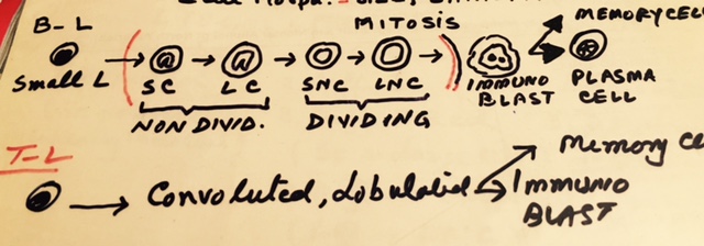
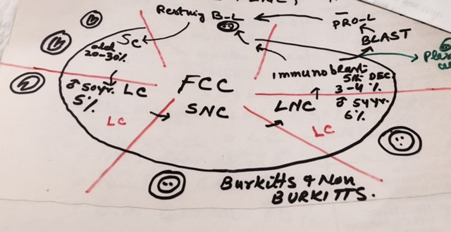
Following diagram shows the differentiation of the cells when stimulated by antigenic stimulation.

Table showing Working Formulation
| Low grade | Intermediate grade | High grade |
| 5 years survival 50 to 70% | 5 years survival 35 to 40% | 5 years survival 23 to 32% |
| 10 years survival 45% | 10 years survival 26% | 10 years survival 23% |
| Small lymphocytes B-L (SLL) | Follicular predominantly large cells (LC + LNC) | Large cells Immunoblastic cells (B and T cells origen) |
| Follicular predominantly small cleaved (SC) | Diffuse small cells (SC) | Lymphoblastic cells |
| Follicular mixed (SC + large cells) | Diffuse mixed cells (SC + large cells ) | Small non-cleaved (Burkitt’s and non-Burkitt’s) |
| Diffuse large cells (LC + LNC ) | Miscellaneous (Mycosis Fungoides, Sezary syndrome, Histiocytosis and HTLV-leukemia) |
LOw Grade Lymphomas
Small lymphocytic lymphoma (SLL)
- These are 4% of the lymphomas.
- These are well differentiated lymphocytic non-hodgkin’s lymphoma.
- These are old age group lymphomas. 6th to 7th decadesis common.
- There is generalized enlargement of the lymph nodes.
- There is mild enlargement of liver and spleen.
- These lymphomas have indolent course, but difficult to eradicate owing to low proliferative index.
- There is no follicular pattern and these are always diffuse .
- These are B – Lymphocytes (95%) with surface marker of IgM, IgD, and CD19+.
- While T – Lymphocytes are 5% in origen.
- There is compact nucleus and mitosis are rare.
- 40 to 60% transform into chronic lymphocytic lymphoma.
- Bone marrow involvement is common.
- Some of these lymphomas produced IgM is called Waldenstrom’s Macroglobulinemia.
- There is prolonged survival .
Follicular Lymphoma
These are 40% of the adult Non-Hodgkin’s lymphoma.
There are types of :
- Small Cleaved cell
- Mixed cells type consists of Small cleaved, Large cleaved and Large non-cleaved.
Follicular Small cleaved cell Lymphoma
- These are most common. These are 60% of the adult Non-Hodgkin’s lymphoma.
- This more common in the middle aged and the elderly people.
- Nucleoli are indistinct and mitosis are rare.
- There are Large cleaved and Large non-cleaved cells are present but these are <20%.
- Median survival is 10 years.
Follicular Mixed Cell Lymphoma
- These are more common in the older age group (50 to 60 years).
- Male, female ratio is equal.
- There is painless enlargement of the lymph nodes.
- Bone marrow involvement is common, seen in 75% of the cases.
- These are >20% and <50% are large cells. There are large cleaved and large non-cleaved cells.
- Transition to diffuse form takes roughly 8 years.
- There are >20% and <50% of the large cells.
- Lymphoma cells show surface markers of Ig, and CD10, CD20 and CD21.
- Median survival is 7 to 9 years.
- Some believes that hands off.
Intermediate Grade Lymphoma
Follicular Predominantly large cells.
- These are uncommon lymphomas <15%.
- There are increased mitosis.
- They change into diffuse form.
- Prognosis is poor because they early transform into diffuse .
- Majority of the large cells are large cleaved and large non-cleaved.
Diffuse Small Cleaved cells lymphoma
- Age is around 60 years.
- There is increase male to female ratio.
- There are increased mitosis.
- Nucleoli are indistinct and coarse chromatin.
- Surface Ig and pan B- antigen (CD19 and CD20) are positive.
- Rarely CD10 may positive.
- Prognosis is poor and median survival is 3 to 4 years.
Diffuse Mixed Cell Type Lymphoma
This lymphoma consists of Small cleaved (SC), Large cleaved (LC), and large non-cleaved (LNC).
Large cleaved cells are larger than normal histiocytes or endothelial cells.
Chromatin is dispersed and nucleoli are indistinct.
Large non cleaved cells are four times the size of small lymphocytes.
Nucleoli are prominent and are 1 to 2.
Nucleus is vesicular and cytoplasm is pale.
Diffuse large cell Lymphoma
- This consists of large cells and Large non-cleaved cells.
- These are most common.
- There is increased male to female ration.
- Median age is 60 years.
- Extranodal signs and symptoms are more common. There is involvement of skin, gastrointestinal tract, bone and brain.
- There is involvement of waldeyer’s ring in more than 50% of the cases.
- There may be diffuse masses in liver, spleen.
- Aggressive chemotherapy is needed.
- Remission may be seen in 60 to 80%.
- 50% of the patient may be symptoms free.
- 70% of the cases there are B- lymphocytes and 30% has T- lymphocyte.
High Grade Lymphoma
These are aggressive lymphomas. Often cured by chemotherapy.
There is high proliferative index which make more susceptible to cytotoxic drugs.
Small Noncleaved Non Hodgkin.s lymphoma are Burkitt’s and Non Burkitt’s lymphoma .
Large Cell Immunoblastic lymphoma
There are wide morphologic changes.
B – Immunoblastic Lymphoma:
- There the cells are 4 to 5 times of the small lymphocytes.
- There is vesicular nuclei and 1 to 2 nucleoli.
- Cytoplasm is deep staining.
- Surface marker is CD19 and CD20 positive.
- 50% has history of autoimmune diseases like Sjogren’s syndrome and Hashimoto’s thyroiditis and AIDs.
T – Immunoblastic lymphoma:
- These are seen in 5th decades.
- These are more common in the male.
- The cells have clear cytoplasm and polymorphous nuclei.
- These represents 20% of the childhood lymphoma.
Lymphoblastic Lymphoma:
- These are most common before the age of 20 years.
- Male : female ratio is 2 : 1.
- In children 40% Non Hodgkin’s lymphoma falls in this group.
- There is early bone marrow , and meninges involvement.
- There may be leukemic phase looks like Acute lymphocytic leukemia.
- There are T – lymphocytes and looks like T- L acute lymphocytic leukemia.
- Cells have uniform and scanty cytoplasm.
- Nucleus is lobulated and nucleoli not prominent.
- Increase mitosis give rise to starry sky appearance.
- Surface markers are CD2, Cd5, CD7 are positive. Some cells show Cd4 and CD8 positive. CD3 is positive/negative.
- Treatment is judt like acute lymphocytic leukemia (ALL) . Aggressive treatment is effective.
