Estrogen/Progesterone Receptors (ER/PR Receptors)
Estrogen/Progesterone Receptors
What samples are needed for Estrogen/Progesterone Receptors?
- This procedure is done on the paraffin blocks of Breast cancer cases.
- This can also be done on fresh biopsy tissue. Place the tissue on ice or in formalin.
- Controls are a must and need to run.
What are the Indications for Estrogen/Progesterone Receptors?
- Estrogen/progesterone receptors are done on breast cancer to find hormone sensitivity and therapy.
- ER/PR is used as a prognostic factor.
What precautions are needed for Estrogen/Progesterone Receptors?
- Delayed fixation may cause deterioration of the receptors and produce a lower result.
- Hormones should be discontinued before the biopsy.
- Tamoxifen therapy may cause a false-negative result.
- Contraceptives or menopausal estrogen, when given, lower the result.
How will you discuss the Pathophysiology of Estrogen/Progesterone Receptors?
- Biochemical concept:
- ER/PR receptors are cellular proteins with high affinity and great specificity for these hormones.
- These receptor proteins are present in other target organs like the uterus, pituitary gland, hypothalamus, and breast.
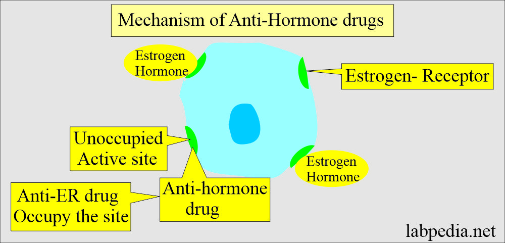
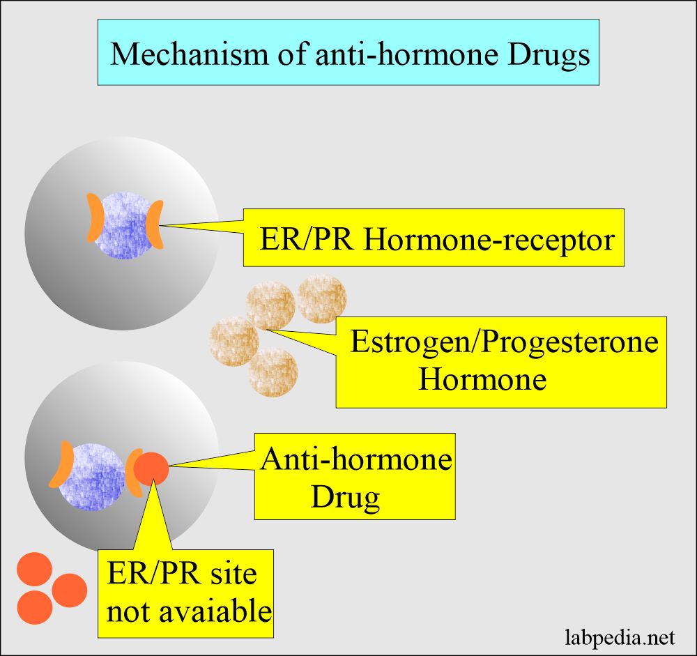
- Estrogen stimulates the biochemical process in the target cells that normally contain estrogen receptors; a reduction in blood estrogen level is expected to reduce the biochemical activity of these cells.
- This is the accepted rationale for using endocrine therapy in females with breast carcinoma.
Discuss ER/PR response to anti-hormone therapy?
- ER/PR are the biomarkers of breast cancers. These can be prognostic, predictive (response to the therapy), or both.
- The higher the ER content positivity, the higher the response rate to endocrine therapy.
- In carcinoma of the breast, 60% of the cases are estrogen receptor-positive.
- Roughly two-thirds of the cases of ER-positive respond to anti-hormone therapy.
- In 95% of the cases, negative ER-receptor fails to respond to anti-hormone therapy.
- Breast cancer ER-positive or PR-positive are twice as sensitive to anti-hormonal therapy (70 % response to anti-hormonal treatment).
- While ER/PR negative cases have a 10% response rate.
- ER/PR-positive cases have more disease-free survival.
- Hormone receptors should be done on all breast cancers.
- ER/PR is more favorable in a menopausal group than in younger patients.
How will you define PR- -receptor?
- It is useful to help in the assay of the ER receptors.
- Metastatic cancer with ER and PR receptor-positive tumors has a response rate of 75% to endocrine therapy.
- If the ER-positive and PR-negative tumors have a 40% response rate.
- The only response rate of ER-negative and PR-positive patients is 25% for endocrine therapy.
- In the case of ER and PR negative, the response rate is only 5%.
- The percentage of positive cases in postmenopausal women is greater than in premenopausal women.
What is the Normal report of ER/PR?
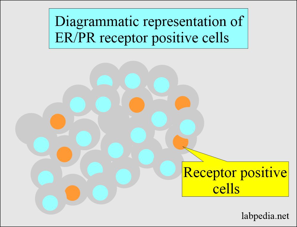
- Negative case = <5 % cell stains for receptors.
- Positive case = >5% of the cells stain for receptors.
Another source
ER/PR receptors assay on tissue:
- Negative = <3 fmol/mg cytosol protein.
- Borderline = 3 to 9 fmol/mg cytosol protein.
- Positive = >10 fmol/mg cytosol protein.
How will describe interpretations of Estrogen/Progesterone Receptors?
- This subjective interpretation depends on the staining intensity and the number of positive cell nuclei.
- Only the cell nuclei staining is considered positive.
- Favorable response >20 % cell stain.
- The borderline response is 11 to 20 % of the cell stain.
- The unfavorable response is < 10 % cell stain.
- ASCO guidelines are:
- Positive for ER/PR if ≥ 1% of the tumor cell nuclei are immunoreactive.
- Negative ER/PR if <1% of tumor cell nuclei are immunoreactive.
- Allred scoring: This replaced the early scoring system.
- ER-positive tumor cells have > 10% positive cells.
- ER-negative tumor cells are 1 to 9% positive cells.
- ER-positive tumor cells have > 10% positive cells.
| Score | Positive cells % | Intensity | Intensity score |
| 0Score | o | None | 0 |
| 1 | None | Week | 1 |
| 2 | 1 to 10 | Intermediate | 2 |
| 3 | 11 to 33 | Strong | 3 |
| 4 | 34 to 66 | ||
| 5 | 66 to 100 |
What is the hormone receptor positivity in different patients?
- ER+ = 80% of the cases.
- ER+ PR+ = 65% of the cases.
- ER+ PR- = 13% of the cases.
- ER- PR+ = 2% of the cases.
- ER- PR- = 25% of the cases.
Response to anti-hormone therapy:
| ER | PR | Response to hormone therapy |
|---|---|---|
| positive | positive | 75 % |
| negative | positive | 60 % |
| positive | negative | 35 % |
| negative | negative | 25 % |
- >50% of the ER-positive cases respond to chemotherapy like tamoxifen, estrogen, androgens, oophorectomy, and adrenalectomy.
- Positivity increases when PR is positive.
- The advantage is that Endocrine, anti-hormone (tamoxifen) therapy is highly effective and relatively non-toxic. It is more tolerable than other modalities like radiation and chemotherapy.
The disadvantage is that ER/PR-negative breast cancer cases have no anti-hormone-positive role.

The brown color indicates a positive reaction (ER/PR+)
Questions and answers:
Question 1: What is the significance of ER/PR positivity?
Question 2: When you will say that ER/PR stain is negative?


