D-Dimer test, Fragment D-dimer, Diagnosis of Disseminated Intravascular Coagulopathy (DIC)
D-Dimer test
What Sample for the D-Dimer test is needed?
- Citrated plasma is needed.
- It is stable for 8 hours at room temp. It can be kept at -20 °C for 6 months.
Indications for D-Dimer test
- This test is done to diagnose DIC (disseminated intravascular coagulopathy).
- It can diagnose other thromboembolic disorders (venous thrombosis).
- It can diagnose Pulmonary embolism.
- For the diagnosis of acute myocardial infarction.
- D-dimer may be increased by atrial fibrillation, congestive heart failure, and cirrhosis.
What is the definition of D-dimer?
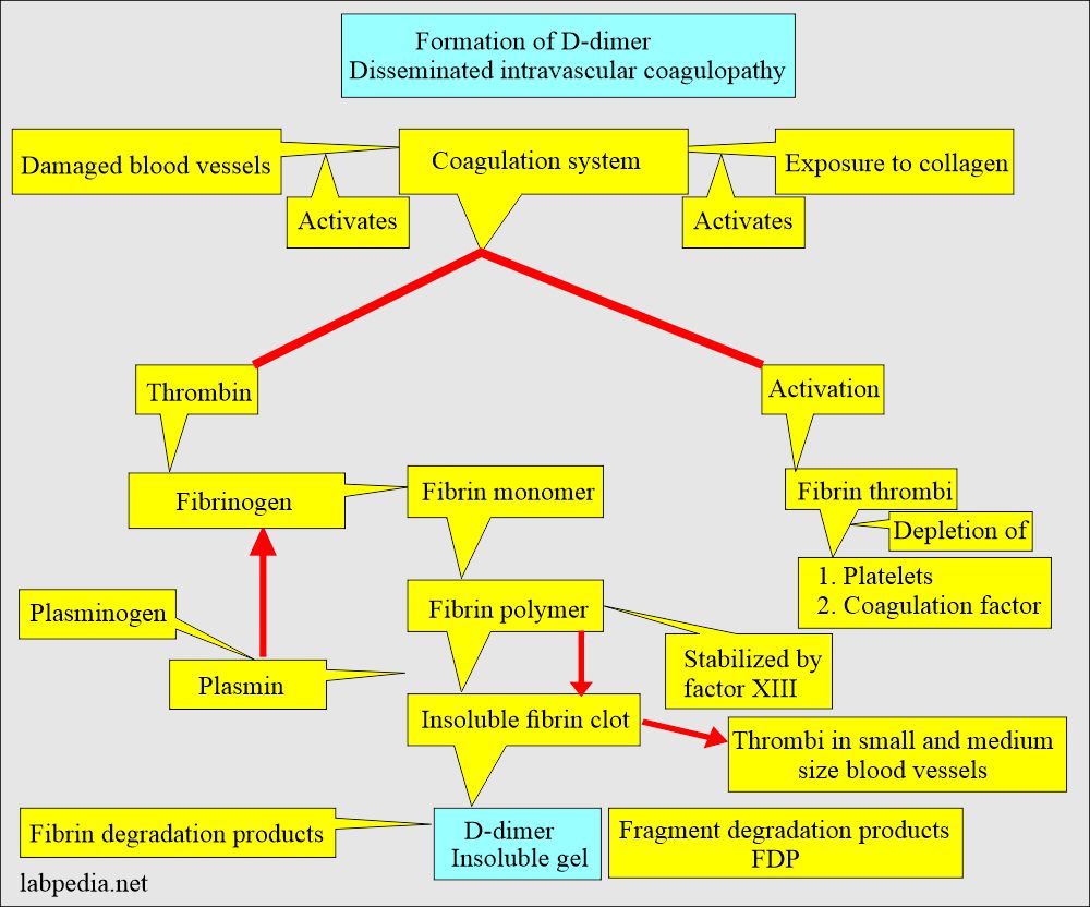
- D-dimers are produced by the action of plasmin on cross-linked fibrin clots.
- The presence of D-Dimer confirms that both thrombin generation and plasmin generation have occurred.
- Factor XIII generates a D-dimer by covalently linking the D regions of the fibrin molecule.
- D-dimer assesses both thrombin and plasmin activity.
- Damaged blood vessels and exposure to collagen activate both intrinsic and extrinsic cascade of blood coagulation.
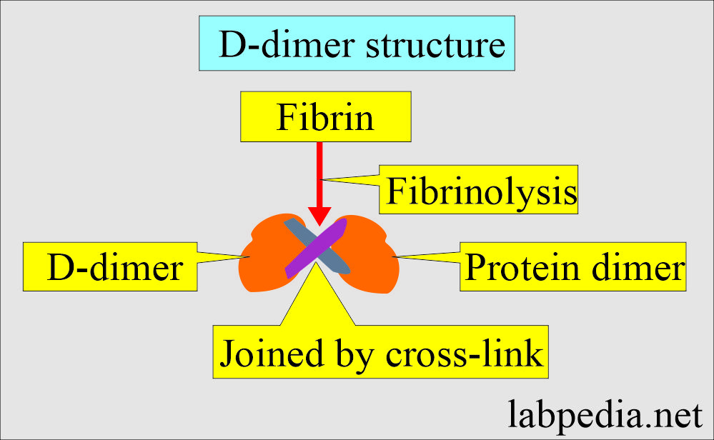
- SO D-dimer is a fibrin degradation product (fragment).
- It is found in the blood as a small protein fragment after the degradation of the fibrin clot (fibrinolysis).
- It consists of 2 D-fragments of fibrin protein joined by a cross-link, so-called protein dimer.
Disseminated intravascular coagulopathy (DIC):
What is the definition of Disseminated Intravascular Coagulopathy (DIC)?
- This is the most serious condition due to fibrinolysis in DIC.
- This is also called consumption coagulopathy.
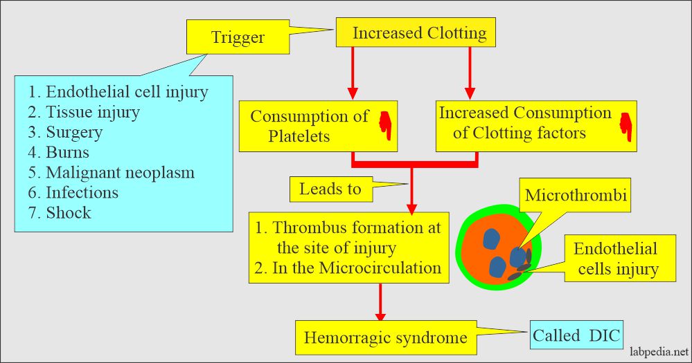
- DIC is an acquired coagulation disorder caused by excessive systemic activation of the coagulation system, showing widespread fibrin thrombi in microcirculation with rapid depletion of platelets and coagulation factors leading to bleeding.
- There is the activation of thrombin leading to thrombosis of small and mid-sized vessels.
- Usually, there is more than one mechanism in action.
- This is a widespread inappropriate intravascular deposition of fibrin with the consumption of coagulation factors and platelets, which occurs due to many diseases with the release of procoagulants into circulation, or this may cause widespread endothelial damage and platelet aggregation.
- There is the deposition of microclots in the blood vessels and depletion of the plasma fibrinogen, leading to hemorrhagic syndrome.
What is the pathogenesis of Disseminated Intravascular Coagulopathy (DIC)?
- This is an acquired clinical syndrome that is due to increased protease activity in the blood.
- It is the unregulated release of thrombin with the formation of fibrin and increased fibrinolysis.
- This is mainly due to the activity of the coagulation cascade (procoagulants).
- The possible factors that will increase the procoagulant’s activity are:
- Arterial hypotension, mostly accompanied by shock.
- Hypoxemia.
- Acidosis (acidemia).
- Stasis of the capillary blood flow.
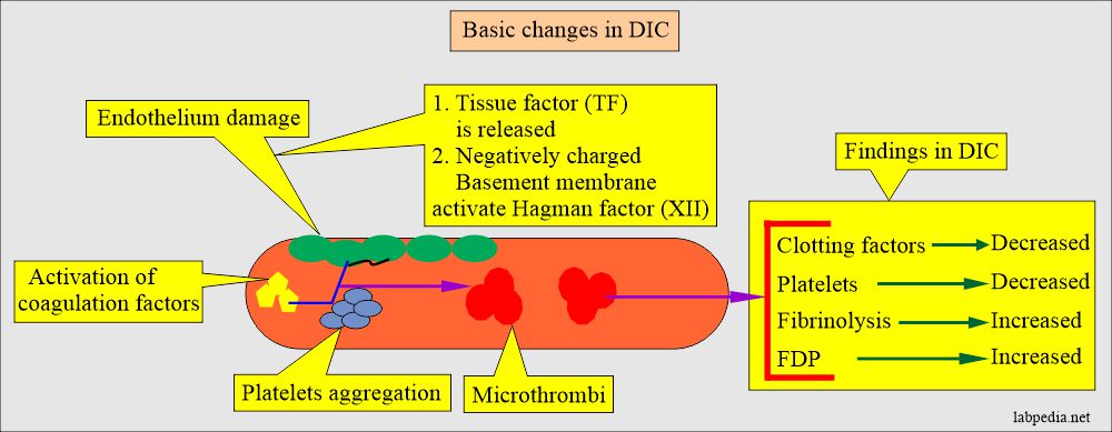
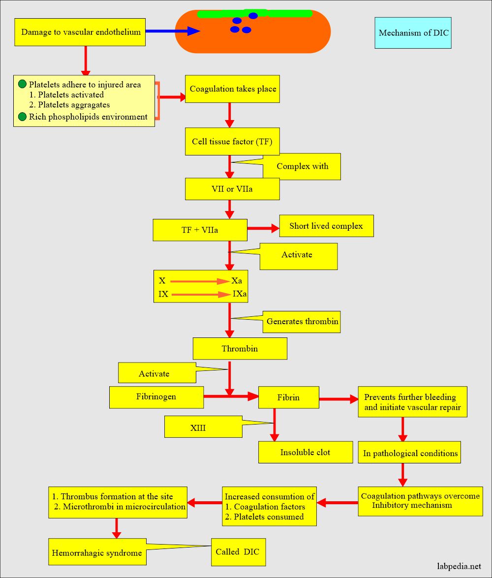
- The DIC is initiated by releasing tissue factor (TF) through direct endothelium damage, activation, or tissue damage.
- Tissue factor (TF) is also released directly into the blood:
- From the white blood cells (monocytes).
- Immune complexes.
- Malignant cells.
- Tissue factor (TF) is also released directly into the blood:
- Tissue factor (TF) is present in endothelial cells. It is released when there is damage to the endothelial cells.
- Cell damaging agents are:
- Endotoxin.
- Cytokines (Interleukin-1, 6, and 8).
- Tumor necrosis factor-α.
- Platelet-activating factor.
- Summary of DIC pathogenesis:
- Initiated by the endothelial cells damage and release of tissue factor (TF).
- The tissue factor (TF) stimulates the coagulation factors.
- Activation of plasmin and thrombin.
- Accelerated consumption of coagulation factors.
- Clotting and hemorrhage will take place.
- Snake venom can activate factor X to Xa.
- Damaged endothelium and inhibited the anticoagulant activity, there is uninhibited thrombin activity and unrestricted formation of clots.
Classification of Disseminated Intravascular Coagulopathy (DIC):
- DIC is divided into two types:
- Acute DIC.
- Chronic DIC.
Acute Disseminated Intravascular Coagulopathy (DIC):
- It is uncompensated DIC.
- Coagulation factors are consumed faster than these are replaced.
- Acute DIC is seen in:
- Trauma.
- Sepsis.
- Shock.
- Burns.
- Eclampsia.
- Amniotic fluid embolism.
- Acute promyelocytic leukemia, FAB M3.
- Lab findings are:
- There is thrombocytopenia.
- Fibrinogen is decreased.
- Factor VIII is decreased.
- There is microangiopathic hemolytic anemia.
- Increased PT, APTT, and positive protamine sulfate test.
Chronic Disseminated Intravascular Coagulopathy (DIC):
- This is compensated by DIC.
- The coagulation factors that are consumed are compensated or replaced.
- Chronic DIC is seen in:
- In various malignancies of the Pancreas, lungs, stomach, and breast.
- It is also seen in leukemias.
- Myeloproliferative disorders.
- Obstetric disorders.
- Paroxysmal nocturnal hemoglobinuria.
- Lab findings are:
- Normal platelet count.
- Normal PT, APTT, and fibrinogenn.
- Positive protamine sulfate test.
| Advised test | Acute DIC | Chronic DIC |
| Platelets count | Decreased (severe) | Decreased (mild) |
| Fibrinogen | Decreased | Increased/normal/moderate decreased |
| Fibrinogen degradation products (FDP) | Positive | Positive |
| Protamine sulfate test | Positive | Positive |
| PT | Increased | Normal/or mild increase |
| Thrombin time | Increased | Normal/or moderate increase |
| APTT | Increased | Normal/or decreased |
| Factor V and VIII | Decreased | Normal |
Mechanism of Disseminated Intravascular Coagulopathy (DIC):
- In DIC, there is increased consumption of the coagulation factors, which will lead to their depletion.
- Also, due to microclots, platelets are depleted.
- Plasmin increases fibrinolysis and leads to fibrinogen degradation products (FDP) and D-dimer formation.
- There is a trigger for the increased clot formation; then fibrinolysis leads to forming FDP and d-dimer.
- Plasmin acts on Fibrin polymer clots and gives rise to FDP and D-dimer.
- FDPs and Plasmin act on the fibrin clot and give rise to D-dimer products.
Causes of DIC are:
- Infections.
- Malignancy.
- Hypersensitivity reactions.
- Vascular abnormalities.
- Widespread tissue damage.
- Other causes like liver failure, pancreatitis, snake venoms, heatstroke, and acute hypoxia.
S/S of DIC are:
- The patient may have weakness and malaise.
- The main feature is bleeding, especially from the site of venipuncture or a recent injury.
- There may be bleeding in the gastrointestinal tract. Oropharynx, into the lungs, and in the urogenital tract.
- During delivery, vaginal bleeding may be very severe.
- Acrocyanosis is irregular-shaped cyanotic patches.
- Microthrombi may cause skin lesions.
- There may be gangrene of the finger or toes.
- There may be occult bleeding to massive GIT bleeding.
- There is abdominal distension.
- Patients may develop renal failure.
- There may be hematuria.
- There is oliguria.
- Patients may have cerebral ischemia.
- There may be subarachnoid hemorrhage.
- There is a slight confusion about convulsions and coma.
- These patients have respiratory symptoms like:
- Cyanosis.
- Hypoxemia.
- Adult respiratory distress syndrome (ARDS).
- Tachypnea.
- Pulmonary infarction.
- Some patients with mucin-producing adenocarcinoma may develop subacute or chronic DIC.
Complications of Disseminated Intravascular Coagulopathy (DIC):
- Thrombotic problems such as deep vein thrombosis, sickle cell anemia, pulmonary embolism, and thrombosis of malignancy are associated with raised levels of D-dimer.
- FDP and d-Dimer form as a complication of disseminated intravascular coagulopathy (DIC).
How do you determine the Diagnosis of Disseminated Intravascular Coagulopathy (DIC)?
- Fibrinogen concentration is low.
- Platelets are low.
- Thrombin time is prolonged.
- Fibrinogen degradation products (FDP) like D-dimer are increased and found in the urine and serum.
- PT and APTT are prolonged in the acute phase.
- The D-Dimer test detects the cross-linked fibrin degradation products.
- The D-dimer assay is more specific than the FDP assay.
- D-dimer is a highly specific measurement of fibrin degradation products that occurs.
- Combining the positive test of FDP and D-dimer is highly specific and sensitive for diagnosing DIC.
- A positive D-dimer result is evidence of DIC or other causes of intravascular thrombosis.
- D-dimer can be used as a screening test for deep vein thrombosis (DVT).
- The fibrin breakdown product concentration will increase when fresh venous thrombosis occurs.
- A negative result is useful in the emergency department for excluding DVT in clinically suspected patients.
Advantages of D-dimer:
- There is no interference from the fibrinogen, so the test can be done on the plasma without any need to collect it in the special tube.
The disadvantage of D-dimer:
- It has limited usefulness because of its use in fibrinolysis and will not detect breakdown products from the fibrinogen.
- D-dimer elevated level has limited value in cancers, inflammation after surgery, or trauma, limiting its usefulness.
The main features of Disseminated Intravascular Coagulopathy (DIC) are due to different factors:
| The groups of Disseminated Intravascular Coagulopathy (DIC) | Main lab criteria |
|
|
|
|
|
|
|
|
What are the Normal values of D-dimer?
Source 1
- Negative
- <0.25 mg/L
Source 2
- Normal = negative ( no D-dimer fragments are seen).
- <250 ng/mL (<250 µg/L).
Source 4
- Normally, plasma is negative for D-dimer.
- Qualitative: It is negative
- Quantitative : < 250 ng/mL or < 250 µg/L ( SI unit)
- Critical value >40 mg/L (40 µg/mL).
Source 6
- <0.4 mcg/mL
What factors will lead to a False-positive test?
- This is seen in heparin therapy.
- The Rheumatoid factor can give false high values of FDP.
- This test is positive in patients after surgery or trauma.
- The false-positive test is seen in estrogen therapy and pregnancy.
Increased Level of D-dimer is seen in the following:
- Primary and secondary fibrinolysis.
- DIC
- Thrombolytic therapy.
- Deep vein thrombosis.
- Pulmonary embolism.
- Arterial thromboembolism.
- Vaso-occlusive crises of sickle cell anemia.
- Pregnancy (Especially the postpartum period).
- Malignancy.
- Surgery.
Questions and answers:
Question 1: What are the complications of DIC?
Question 2: What is lab diagnosis of Disseminated Intravascular Coagulopathy (DIC) ?
- Please see more details in the fibrinogen degradation products FDP.


very informative. Do visit our website https://medintu.in/d-dimer-test/vizag/
Your website looks very good. My website is my hobby and charity.
Really very nice article. thanks for sharing. check this also- https://www.deepnursingbureau.com/
Thanks.