Complete blood count (CBC):- Part 2 – Red Blood Cells Morphology, Interpretations and Hemoglobin Formation
Red Blood Cells Morphology
What sample is needed for Red Blood cell morphology?
- Takes blood in the EDTA.
- Direct blood smears for the appreciation of the RBC morphology.
What are the indications for a Complete blood count (CBC)?
- To diagnose the various types of anemia.
- Any other abnormality of white blood cells and platelets.
What is the structure of the Red blood cell membrane?
- Red blood cell metabolism is important for its survival and function.
- The RBC membrane consists of proteins and phospholipids.
- It can change its shape without any fragmentation or damage.
- The RBC membrane comprises:
- 50% proteins.
- 20% phospholipids.
- 20% cholesterol.
- 10% carbohydrates.
- Carbohydrates are only in the outer layer.

Red Blood Cells Morphology: RBC cell membrane and functions

Red Blood Cells Morphology: RBC structure

RBC structure
Hemoglobin formation, structure, and functions:
What is the structure of Hemoglobin?
- Normal hemoglobin formation is dependent upon:
- Adequate iron supply and delivery.
- Adequate formation of the precursor of the heme, protoporphyrin.
- Adequate synthesis of globin.
- Hemoglobin (Hb) is a conjugated globular protein with a molecular weight of around 64.4 kDa.
- This protein is 95% of the RBC’s dry weight or 33% of the RBC’s weight by volume.
- Around 65% of the Hb synthesis occurs during the nucleated stage of RBC maturation, and 35% occurs during the reticulocyte stage.
- Hemoglobin genes are present on chromosomes 16 and 11.

Hemoglobin gene’s location
- Normal Hemoglobin consists of two α-chains and two β-chains with four protoporphyrin rings with Fe++ (ferrous).
 normal structure")
Hemoglobin (Hb) normal structure
What are the functions of hemoglobin?
- Hemoglobin is the main oxygen carrier to the cells and tissues.
- This also takes CO2 back to the lungs.
What are the energy pathways for RBC metabolism?
- Active RBC metabolic pathways are important for the production of an adequate amount of ATP, which is necessary for:
- Hemoglobin function.
- RBC membrane integrity and change in shape.
- An adequate amount of reduced pyridine nucleotides.
- Maintaining the RBC volume.
- RBC generates energy mainly from the anaerobic breakdown of glucose.
- Mature RBC has a limited ability to metabolize fats and amino acids.
- RBC ATP needs 90% of its energy generated by the Embden-Meyerhof glycolytic pathway.
- 5% to 10% of the energy is provided by the metabolism of the glucose-hexose monophosphate shunt.

RBC source of energy mechanism
What is the RBC structure and life pattern?
- RBCs from the front side are round but appear biconcave when viewed from the side.
- This can be illustrated by a doughnut-shaped object with a depression in the center.

RBC morphology
- It measures 7 to 8 µm in diameter.
- RBCs mature in the bone marrow, and when they come to the peripheral blood, it takes 3 to 5 days.
- The RBCs live in the peripheral blood for around 120 days. After completing their life, these are cleared in the spleen, liver, and bone marrow.
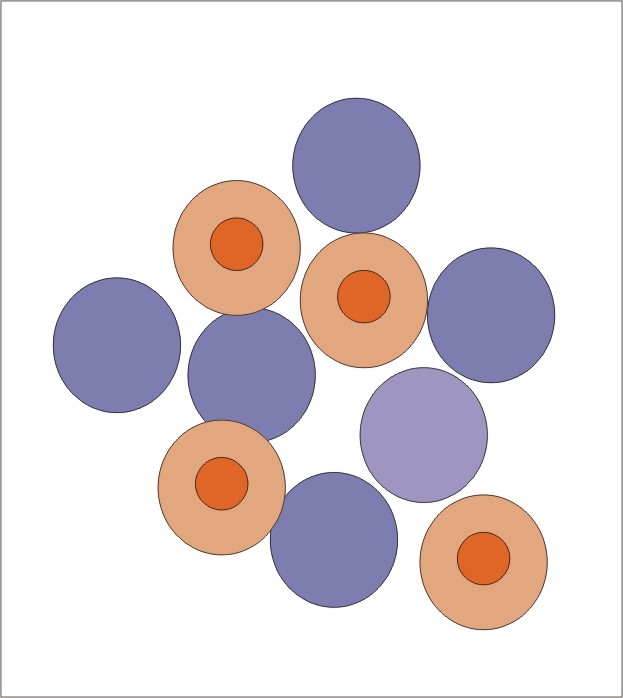
What are the abnormal forms of the red blood cells (RBCs)?

Various forms of RBCs
What are the functions of the red blood cells?
- The RBCs’ primary function is to carry oxygen from the lungs (arterial blood) to the cells and tissues. The oxygen is carried by a chemical combination with hemoglobin.
- At the tissue level, oxygen is exchanged with CO2, bringing CO2 in venous blood to the lungs.
- Normally, O2 exchange occurs between 95% saturation in arterial blood with a mean arterial O2 tension of 95 mmHg and 70% saturation in venous blood with a mean venous O2 tension of 40 mmHg.

RBC role in oxygen transport
: Hemoglobin role in Oxygen transport")
Complete blood count (CBC): Hb role in oxygen transport
- The RBCs must be in close contact with the tissue and have successful gaseous exchange to carry hemoglobin. RBCs must be able to pass through the microcirculation.
- RBCs travel in the microcirculation in 120 days; roughly, this covers approximately 300 miles (480 km).
- The RBCs are flexible and biconcave discs that fulfill all the above functions.
What are the clinical interpretations of the Peripheral blood smears?
- It provides extensive information:
- RBC morphology.
- Effects of various drugs.
- Different types of anemias.
What is the Anemia workup?
- Hb concentration.
- Hematocrit (Hct).
- RBC indices.
- Reticulocyte count.
- Evaluation of the peripheral blood smear.
- Bone marrow can also be advised to classify anemia.
What are the defects of the RBC formation and the reason for anemia (Etiology)?
- Deficiency diseases.
- Refractory anemia due to ineffective erythropoiesis.
- Defect of the bone marrow due to hypoproliferative anemias.
- Excessive loss of blood, such as:
- Hemolysis.
- Hemorrhage.
What are the causes of the RBCs leading to various types of anemias?
- Iron-deficiency anemia.
- Sideroblastic anemia.
- Anemia of chronic diseases.
- Thalassemia syndrome.
- Pernicious anemia.
- Folic acid deficiency anemia.
- Aplastic anemia.
- Pure red cell aplasia.
- Refractory anemia.
- Paroxysmal nocturnal hemoglobinuria.
- Hemolytic anemias.
- Hemorrhagic anemia.
- Hereditary spherocytosis.
- Hereditary elliptocytosis.
- Glucose-6-phosphate dehydrogenase deficiency.
- Pyruvate kinase deficiency.
- Sickle cell anemia and disease.
- Autoimmune hemolytic anemia.
What are the bases for assessing red blood cells?
RBC volume:
- Normal MCV, RBCs are called normocytic.
- High MCV indicates that RBCs are macrocytic.
- A low MCV indicates that RBCs are microcytic.
Hemoglobin contents:
- Normal Hb and MCHC are called normochromic.
- High MCHC RBCs are called hyperchromic.
- Low MCHC RBCs are called hypochromic.
How will you summarize various anemias?
| Clinical presentation | MCV fl |
MCH pg |
MCHC % |
|
|
|
|
|
|
|
|
|
|
|
|
|
|
|
|
Microscopic evaluation of peripheral blood smear:
What information can we obtain from a peripheral blood smear?:
Microscopic examination of the peripheral blood smears gives information about:
- The size (anisocytosis).
- The shape (poikilocytosis).
- The color (hypochromic or hyperchromic).
- Intracellular inclusions.
How will you assess the microscopic examination of a peripheral blood smear?
- First scan under the low power (10x) and note:
- Staining quality of the smear.
- Also, check the center and edges of the slide to ensure there is no clumping of RBCs, WBCs, and platelets, indicating good cell distribution on the smear.
- Scan for abnormal cells.
- Determine the area where the RBCs should not overlap.
- The RBCs should have a gradual central pallor.
- Scan the smear in high power (40x) and note:
- WBCs abnormality.
- Can estimate the WBCs count.
- Evaluate the RBC morphology.
- Scan the smear in oil immersion (100x) and note:
- Evaluate the RBC’s anisocytosis, poikilocytosis, hypochromasia, polychromasia, and inclusion.
- Can evaluate the platelet count.
- WBCs differential can be done.
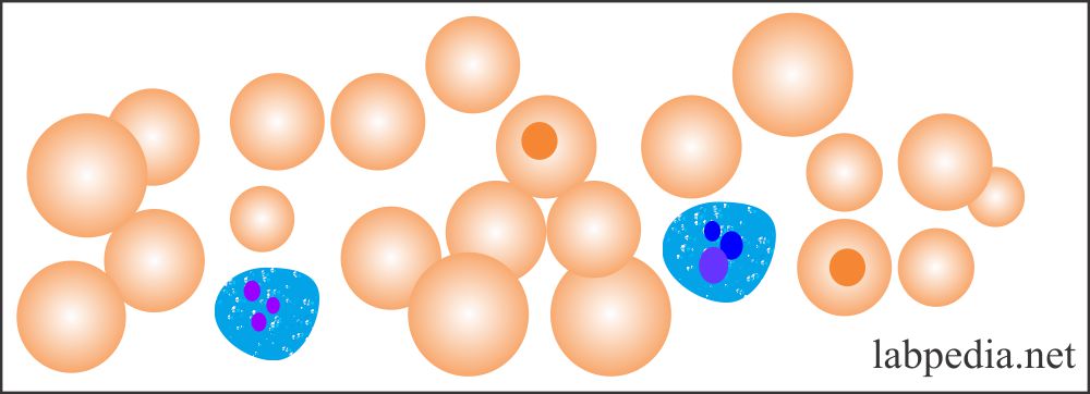
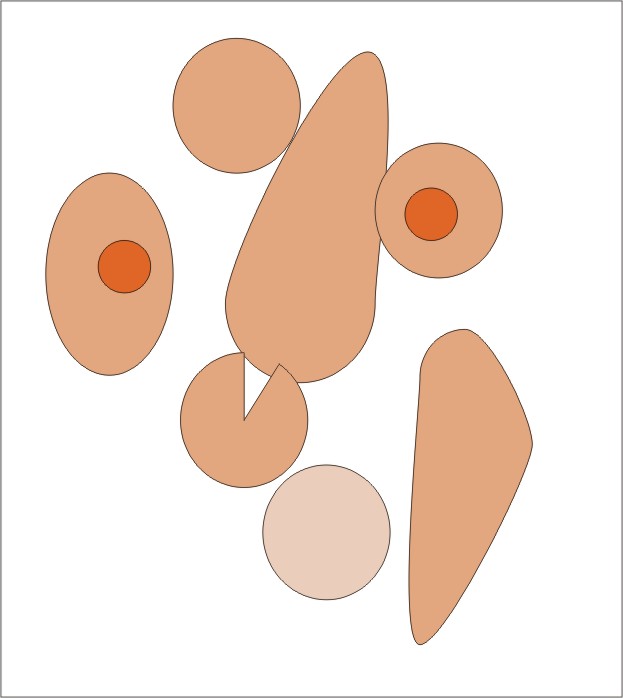
How will you summarize the RBCs’ morphology changes and interpretation?
| Terminology | Morphology | Explanation | Interpretation (Causes) |
|
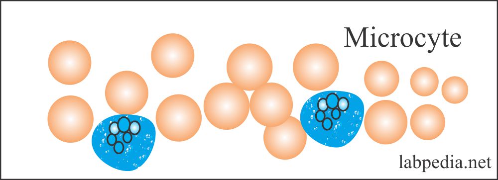 |
RBC size is smaller than normal |
|
|
 |
RBC size is larger than normal |
|
|
 |
Variation in RBC size |
|
|
 |
Variations in RBC shapes |
|
|
|
RBCs are large and basophilic. |
|
|
|
RBCs are small and round and have no central pale area. | Seen in:
|
|
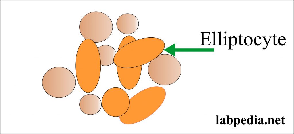 |
RBCs are rod-shaped (oval or elliptical) |
|
|
 |
RBCs have a darkly central area. |
|
|
 |
RBC looks like tears |
|
|
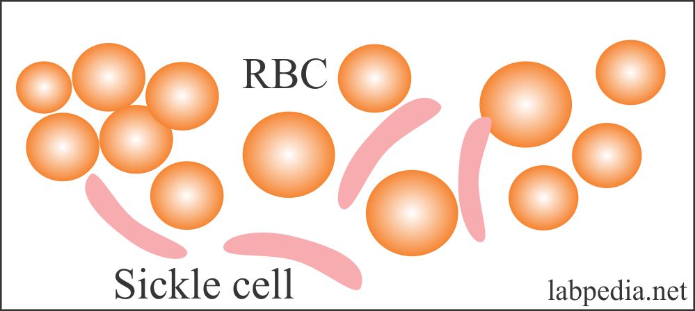 Sickle cell |
Moon-shaped RBC |
|
|
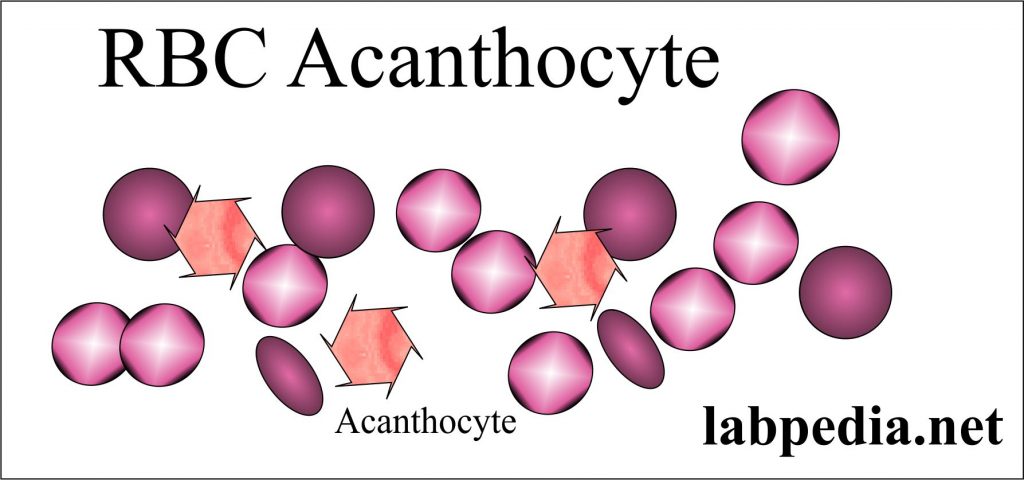 |
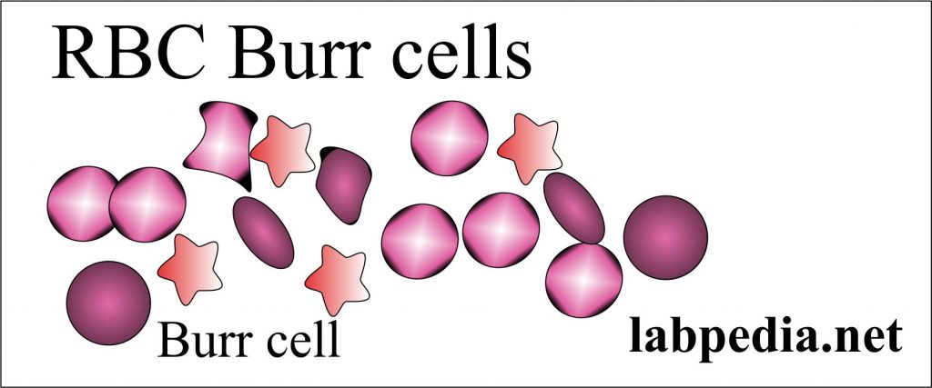
RBCs have spikes, irregular projections |
|
|
 |
RBCs show fragments and a variety of shapes and sizes. |
|
|
 |
Elongated RBC |
|
|
 |
RBCs are created and irregular. | Seen in:
|
|
|
RBCs show nuclear remnants. |
|
|
RBCs show denatured hemoglobin. |
|
|
|
Increased granules in polys |
|
|
|
 |
RBCs show clumping or aggregation. | Seen in:
|
|
 |
RBCs are clumped |
|

Red Blood Cells Morphology: Red blood cell morphology
Questions and answers:
Question 1: What is the criterion for the macrocytes?
Question 2: When will you see Howel-jolly bodies in RBCs?