HealthFlex
×
  • Home
  • Immunology Book
  • Lab Tests
    • Hematology
    • Fluid analysis
    • CSF
    • Urine Analysis
    • Chemical pathology
    • Blood banking
    • Fungi
    • Immune system
    • Microbiology
    • Parasitology
    • Pathology
    • Tumor marker
    • Virology
    • Cytology
  • Lectures
    • Bacteriology
    • Liver
    • Lymph node
    • Mycology
    • Virology
  • Blog
    • Economics and technical
    • Fitness health
    • Mental health
    • Nutrition
    • Travel
    • Preventive health
    • Nature and photos
    • General topic
  • Medical Dictionary
  • About Us
  • Contact

Cerebrospinal fluid Xanthochromia (CSF)

August 29, 2023CSFLab Tests

Cerebrospinal fluid Xanthochromia

Specimen for Cerebrospinal fluid Xanthochromia

  • Cerebrospinal fluid is needed.

Indication

  • To assess the presence of increased protein in the CSF.

 Xanthochromia:

Definition of Xanthochromasia:

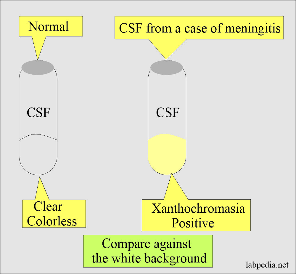
  1. Xanthochromia is a yellow color, and it may occur due to hemoglobin pigments from lysed RBCs, or it appears when CSF proteins are >150 mg/dL and produce a faint yellow color.
  2. Severe jaundice may produce discoloration that may simulate xanthochromasia.

Pathophysiology of Cerebrospinal fluid (CSF):

  1. The normal color of the CSF is clear and colorless.
  2. The color appearance depends upon the presence of hyperbilirubinemia, hypercarotenemia,  melanoma, and elevated protein levels.
  3. A cloudy appearance indicates an increased number of WBCs or increased proteins.
  4. A red tinge indicates blood that may be due to hemorrhage in the subarachnoid space or due to a traumatic tap.
  5. Due to traumatic tap, blood may form clots, which will not occur in the subarachnoid bleeding.
    1. In traumatic tap, successive tubes become clear of blood.
  6. Xanthochromia is usually referred to as a yellow tinge.

How to check xanthochromia:

  • The easiest way is to centrifuge the CSF and see it against the white background.
Cerebrospinal fluid Xanthochromia: CSF Xanthochromia

CSF Xanthochromasia

  • Result
    Xanthochromia, when positive in the CSF, indicates a raised protein level.
  • Please see more details in the CSF fluid analysis.

Possible References Used
Go Back to CSF

Add Comment Cancel


The reCAPTCHA verification period has expired. Please reload the page.

  • Lab Tests
    • Blood banking
    • Chemical pathology
    • CSF
    • Cytology
    • Fluid analysis
    • Fungi
    • Hematology
    • Immune system
    • Microbiology
    • Parasitology
    • Pathology
    • Tumor marker
    • Urine Analysis
    • Virology

About Us

Labpedia.net is non-profit health information resource. All informations are useful for doctors, lab technicians, nurses, and paramedical staff. All the tests include details about the sampling, normal values, precautions, pathophysiology, and interpretation.

[email protected]

Quick Links

  • Blog
  • About Us
  • Contact
  • Disclaimer

Our Team

Professor Dr. Riaz Ahmad Bhutta

Dr. Naheed Afroz Syed

Dr. Asad Ahmad, M.D.

Dr. Shehpar Khan, M.D.

Copyright © 2014 - 2025. All Rights Reserved.
Web development by Farhan Ahmad.