Anti-parietal cell antibody (APCA)
Anti-parietal cell antibody (APCA)
What sample is needed for Anti-parietal cell antibody (APCA)?
- Venous blood is needed to prepare the serum.
- The patient’s serum is needed and should be stored at -20 °C.
- How to get good serum: Take 3 to 5 ml of blood in a disposable syringe or a vacutainer. Keep the syringe for 15 to 30 minutes at 37 °C and then centrifuge for 2 to 4 minutes to get the clear serum.
What are the indications for Anti-parietal cell antibody (APCA)?
- This antibody test is done to diagnose the autoimmune type of Pernicious anemia.
What are the precautions for Anti-parietal cell antibody (APCA)?
- APCA is present in many healthy persons over the age of 60 years.
How will you discuss the pathophysiology of Anti-parietal cell antibody (APCA)?
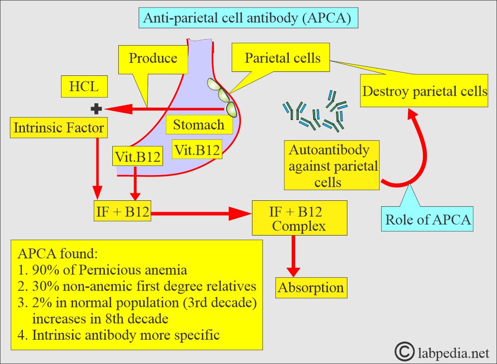
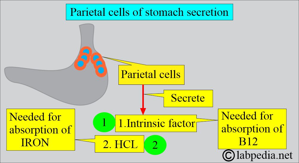
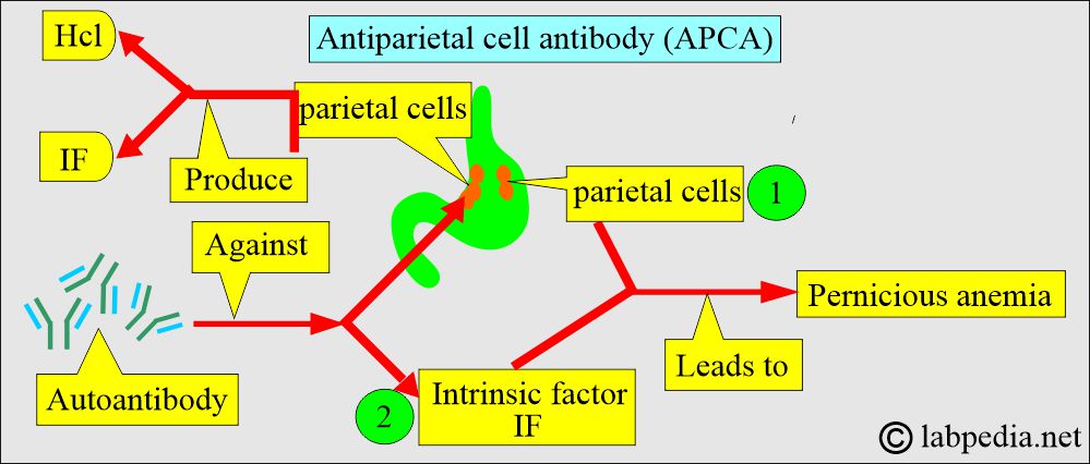
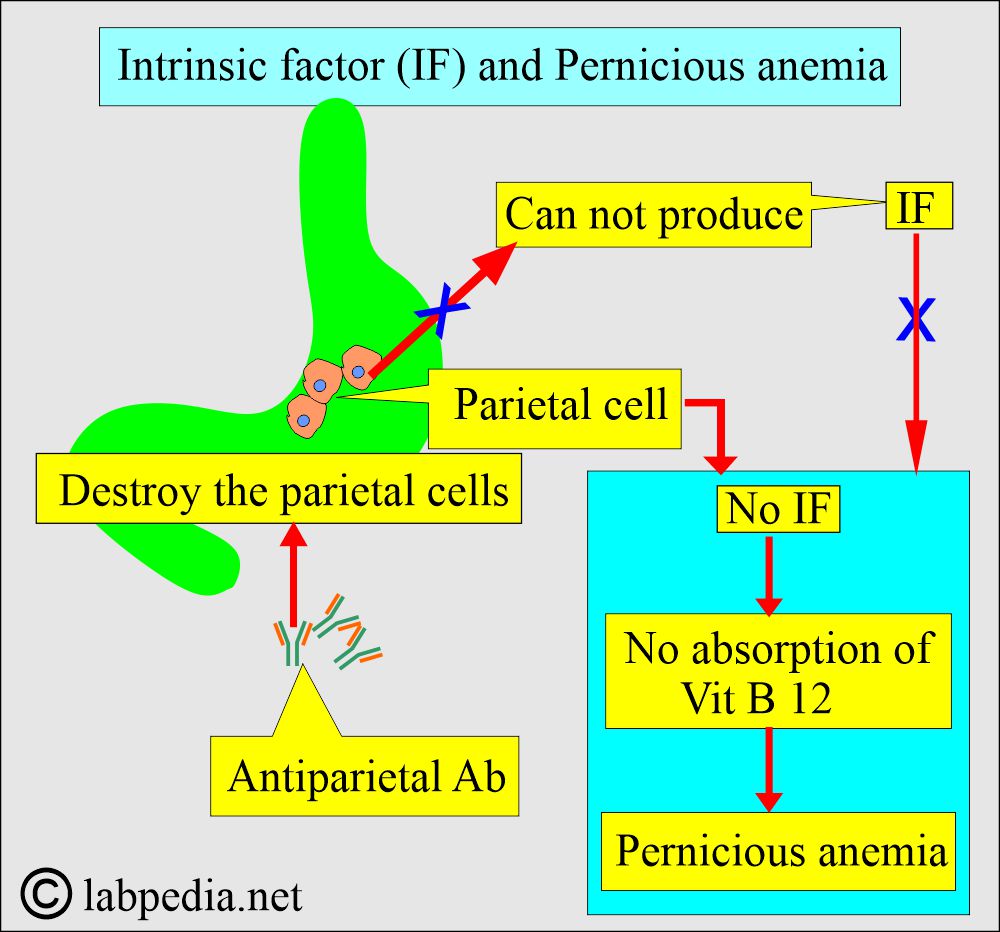
- The parietal cells are present in the proximal portion of the gastric mucosa and produce acid (HCL) and Intrinsic factors.
- The intrinsic factor is needed for the absorption of vit B12.
- A lack of an intrinsic factor due to the antiparietal cell antibodies (APCA) will lead to pernicious anemia.
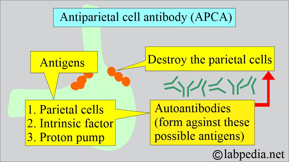
- These antiparietal cell antibodies lead to the destruction of the gastric mucosa.
- Parietal cell antibodies occur in patients with pernicious anemia (50% to 100%).
- There will be a disruption (no production) of the intrinsic factor by the parietal cells.
- In pernicious anemia, two types of antibodies were found:
- One against the parietal cells.
- Second, against the intrinsic factor.
- This antibody can be seen in other autoimmune diseases (20% to 30%), such as Thyroiditis, myxedema, juvenile diabetes, Addison’s disease, and iron-deficiency anemia.
How will you discuss Anti-parietal cell antibodies (APCA)?
- These are 95% positive in cases of Pernicious anemia (another reference says 76% to 91%).
- The normal population has 10% to 15% of these antibodies.
- This is nonspecific to the IF antibody.
- With increasing age, the incidence of APCA increases, especially in the relatives of pernicious anemia patients.
- In pernicious autoimmune anemia, antiparietal antibodies are >80% positive; 50% have antibodies to intrinsic factors.
- Antiparietal antibodies are seen in healthy adults >60 years of age.
- Sometimes these antiparietal antibodies are seen in:
- Atrophic gastritis (idiopathic atrophic gastritis shows 30% to 60% APCA.
- Gastric ulcer.
- Gastric malignancies.
- Juvenile diabetes mellitus.
- Iron-deficiency anemia.
- Myxedema.
- Thyroiditis.
- Addison’s disease.
- APCA, when positive, then needs a more invasive procedure like a gastric biopsy to rule out a gastrointestinal disease.
What are the other possibilities of APCA in various diseases?
| Anti-parietal cell antibody (APCA) | Positivity is associated with various diseases |
|
|
|
|
|
|
|
|
|
|
|
|
What are the normal antiparietal cell antibodies?
- These are normally negative.
Source 4
- Negative anti-parietal antibody by IFA technique.
- When positive, then titrate the serum.
- Positive = Titer level of 1:240
What are the positive possibilities of Anti-parietal cell antibody (APCA)?
- Pernicious anemia (in 95% of the cases, although its specificity is low).
- Atrophic gastritis (30% to 60%).
- Gastric cancer and gastric ulcer.
- These are present in 25% to 30% of the autoimmune diseases of the thyroid.
- Hashimoto’s thyroiditis (25%).
- Myxedema.
- Thyrotoxicosis (25% to 35%)
- Juvenile diabetes (12% to 28%).
- Addison disease.
- Iron deficiency anemia.
- A normal person (5% to 10%).
What are the causes of increased Anti-parietal cell antibody (APCA)?
- Pernicious anemia.
- Atrophic gastritis.
- Myxedema.
- Hashimoto’s thyroiditis.
- Juvenile diabetes.
- Addison disease.
Questions and answers:
Question 1: Which antibody is specific for pernicious anemia?
Question 2: APCA does positive in atrophic gastritis?

