Tumor Markers:- Part 1 – Tumor Markers, Staging and Grading
Tumor Markers
How will you define Tumor Markers?
- Tumor markers are the biochemical or immunological counterparts of the differentiation state of the tumor.
- Tumor markers represent the expression of the substances produced normally by embryogenically related tissues.
- These are mostly proteins produced by the cancer cells.
- Tumor markers are the substances found in increased concentration in malignancies in:
- Blood.
- Body fluids.
- Body tissues.
- Urine.
- Tumor markers can be measured:
- Qualitatively.
- Quantitatively.
- Tumor markers can be measured by:
- Chemical methods.
- Immunological methods.
- Molecular biological methods.
- An ideal tumor marker should be both specific and sensitive to detect small tumors in the early stages.
- But unlikely this is not the situation.
- These are found in different tumors, even originating from one source.
- These are present in higher concentrations in malignancies than in benign tissue.
What are the advantages of tumor markers?
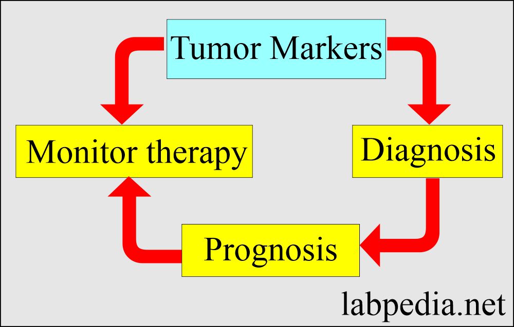
- The tumor markers help diagnose, prognosis, and monitor the tumor.
- Help determine the progression of the disease.
- Help monitor the tumor after treatment.
- It can be used to Screen the general population.
- These are helpful for the Clinical staging of the tumors.
- Helped evaluate the success of the treatment.
- Very useful for the Detection of the recurrence of the tumor.
- Helpful to decide on the immunotherapy.
What are the disadvantages of tumor markers?
- Most of the markers do not differentiate between benign and malignant.
- The cost for the screening of neoplasia is high.
- Tumor markers may not be ideal for screening purposes compared to treatment response.
How will you define the Staging and Grading of the cancer?
- The latest TNM classification was published by the Union for International Cancer Control (UICC) and the American Joint Committee on Cancer (AJCC) in 2017.
How will you discuss the Staging of the tumors?
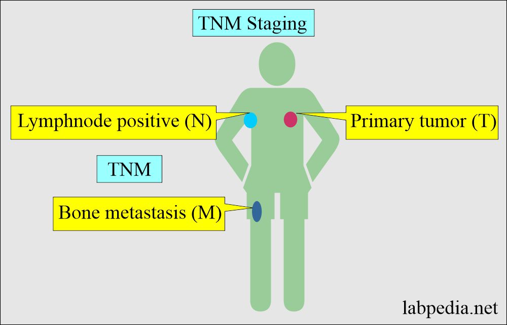
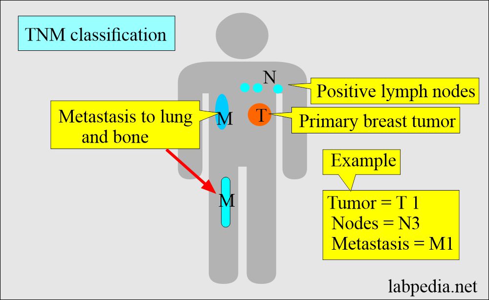
The most common is the TNM classification. This depends upon the spread of the tumor.
- T = Primary tumor size.
- N = Lymph nodal status is the regional lymph node involvement.
- M = Metastasis of the tumor describes the distant spread of the tumor.
Primary tumor size:
- TX = The Main tumor cannot be measured.
- T0 = Main tumor cannot be found.
- T1 = Size and extent of the tumor.
- T2 = same as above
- T3 = same as above
- T4 = same as above. As the T1 to T4, similarly, the tumor size increases.
Lymph node status:
- NX = Cancer in nearby lymph nodes cannot be measured.
- N0 = There is no tumor infiltration in the near regional lymph nodes.
- N1 to N3 = Indicates the number of lymph nodes involved.
Distant metastasis:
- MX = Metastasis can not be measured.
- M0 = No distant metastasis found.
- M1 = Positive for distant metastasis.
How will you discuss the Grading of the tumor?
- This is the histologic appearance of the tumor cells and their abnormal appearance under the microscope.
- This will indicate how the tumor will grow and spread.
This is the histologic differentiation of the tumor. This may be:
- Well-differentiated.
- When the tumor cells and organization are near the normal cells and the tissue.
- These tumor cells grow and spread at a slow rate.
- Moderately differentiated.
- Poorly differentiated. When the tumor cells are abnormal and fast-growing.
- Undifferentiated.
What are the criteria for the grading of tumors?
- Grade 1 = when the tumor cells are near the normal tissue.
- Grade 2 = when the tumor cells are slightly away from the normal tissue.
- Grade 3 = when the tumor cells grow rapidly and faster.
- Grade 4 = when the tumor cells grow more rapidly and abnormal-looking than the normal tissue.
What are the grades of tumors?
- G1= Well-differentiated and low grade.
- G2 = Moderately differentiated and are intermediate grades.
- G3 = Poorly differentiated and high grade.
- G4 = Undifferentiated and a high grade.
- The latest classification given by the College of Americal Pathologists is followed.
What are the objectives of TNM classification?
- This will help in treatment planning.
- This will give an idea about the prognosis.
- This will evaluate the treatment result.
- It will facilitate the exchange of information between the centers.
- It supports cancer control activities, including cancer registries.
- This will contribute to the investigations of human malignancies. (Source = UICC).
Questions and answers:
Question 1: What is the grading of the tumor?
Question 2: What is G4 tumor?


