Serum Protein Electrophoresis, Total protein, albumin and globulin
Serum Protein Electrophoresis
What sample is needed for Serum Protein Electrophoresis?
- This is done on the patient’s serum.
- Also can be done in the urine. 24-hours of urine is preferred.
- CSF for the oligoclonal band.
What are the indications for Serum Protein Electrophoresis?
- This can diagnose some inflammatory diseases.
- It can diagnose neoplastic diseases like multiple myeloma and lymphoma.
- Advised In immune disorders.
- Advised In liver diseases.
- Advised In nephrotic syndrome.
- Advised In Chronic edema.
- To detect monoclonal proteins (Immunoglobulins).
- CSF for the oligoclonal band.
How will you define electrophoresis?
- This is the migration of charged solutes or particles in a liquid medium under the influence of the electrical field.
- Analytes of interest are:
- Proteins.
- Peptides.
- Aminoacids.
- Nucleic acid.
- OLigonucleotides.
- Cations in body fluids and the tissue.
How will you explain the pathophysiology of serum proteins?
- Proteins are constituents of muscles, hormones, hemoglobin, and transport proteins.
- Serum proteins are a source of nutrition and a buffer system.
- Immunoglobulins function in the immune system.
- Carrier proteins, like haptoglobins, prealbumin, and transferrin, transport certain ions and molecules to their site of action.
- Some of the protein regulates:
- The activity of various proteolytic enzymes.
- The osmotic pressure within the vascular compartment.
- Metabolic substances like a hormone.
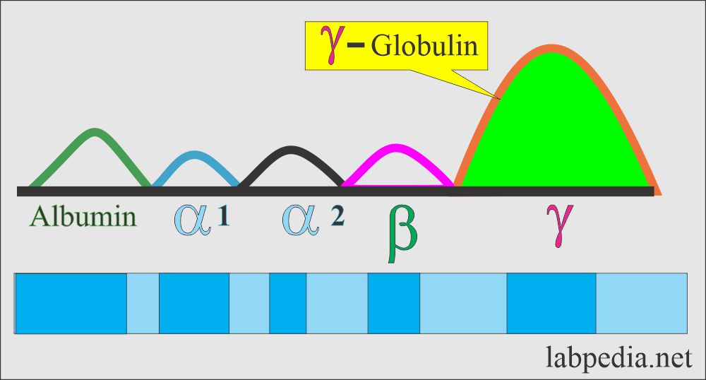
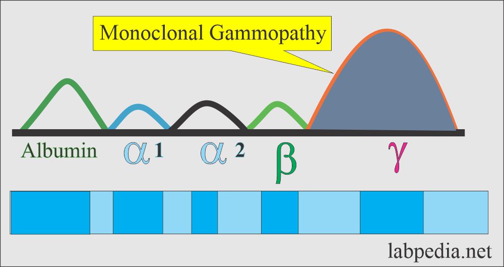
- Immunoglobulins are a major component of gamma-globulins.
- In a normal person, the immunoglobulins are polyclonal.
- The monoclonal band of immunoglobulin indicates a neoplastic process like multiple myeloma and Waldenstrom’s macroglobulinemia.
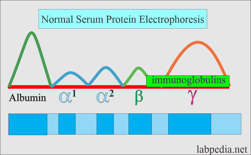
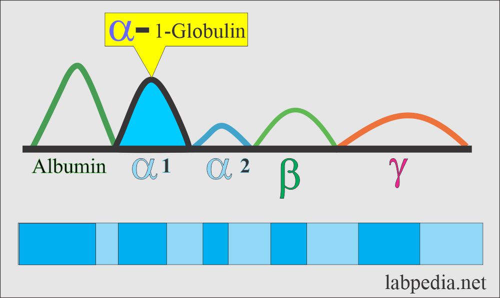
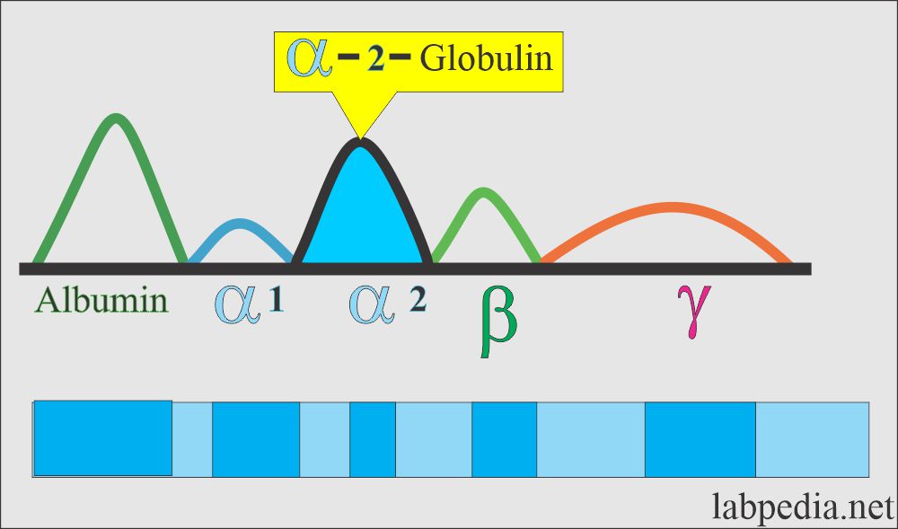
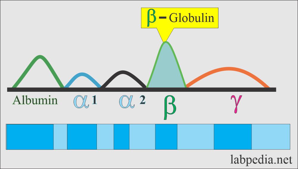
- Serum electrophoresis separates proteins into 5 different bands:
- Albumin, and Prealbumin. Albumin is formed in the liver and is 60% of total proteins.
- alpha1-globulin (α1-globulin).
- alpha 2-globulin (α2-globulin).
- beta – globulin (β-globulin).
- gamma – globulin (γ-globulin).
What is the normal value of serum protein?
| Protein | Adult value g/dL | Cord blood g/dL | Mother’s serum g/dL |
| Albumin | 3.5 to 5.0 | 3.3 | 4.2 |
| α1-Globulin | o.1 to 0.4 | 0.0 | 0.3 |
| α2-Globulin | 0.3 to 0.8 | 0.4 | 1.2 |
| β-Globulin | 0.6 to 1.1 | 0.7 | 1.3 |
| γ-Globulin | 0.5 to 1.7 | 1.0 | 1.3 |
What are the patterns of different zones 0n Serum Protein Electrophoresis?
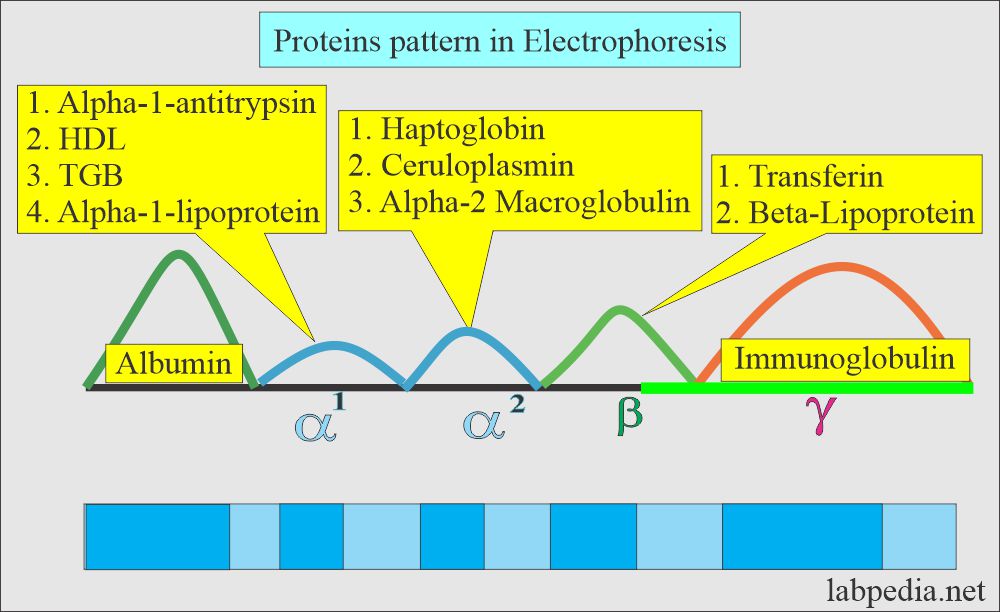
- These different bands have different zones with the characteristic presence of different proteins. e.g.
- The Albumin zone shows only albumin.
- alpha1- zone shows:
- alpha1 – lipoprotein.
- High-density lipoprotein (HDL)
- Alpha-1 -antitrypsin.
- Alpha 2 zone shows:
- Alpha-2 macroglobulins.
- Haptoglobin.
- β -lipoprotein.
- Beta zone shows:
- Transferrin.
- Complement 3 (C3).
- Gamma zone shows
- Fibrinogen.
- IgA.
- IgM.
- IgG.
What are the functions of different proteins?
Albumin :
- Maintain the colloidal osmotic pressure.
- Albumin also transports drugs, hormones, and enzymes.
- Albumin measures liver function. Its concentration is markedly low in liver diseases when liver cell damage occurs.
- The half-life of albumin is 12 to 18 days so that liver cell damage will be observed after this period.
Globulins :
- These are the main components of immunoglobulins (Antibodies).
- Some are transporting proteins like thyroid and cortisol binding proteins.
- Haptoglobin binds hemoglobin during hemolysis.
- Ceruloplasmin is a carrier of copper.
- They may also act as transport vehicles.
What is the Principle of Electrophoresis?
- This is basically the separation or migration of charged solutes of particles in a liquid medium under the influence of the electrical field.
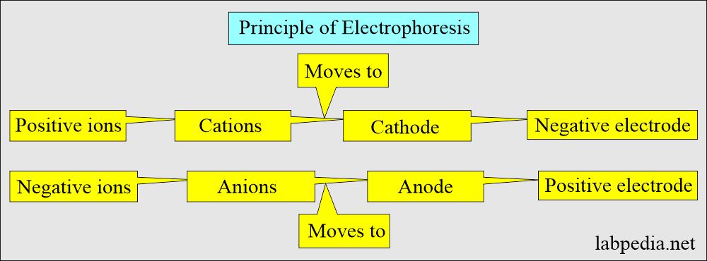
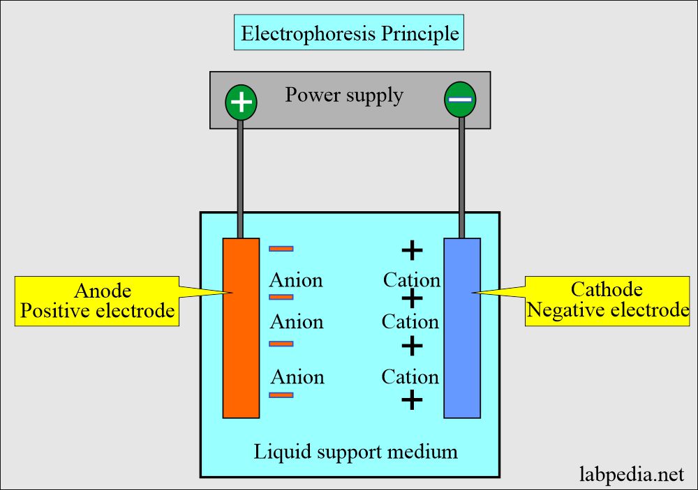
- Chemical substances carrying charges because of ionization move toward either the cathode (Negative electrode) or the anode ( positive electrode). So, protein ions (cation) move toward the cathode, and negative ions (anion) move toward the anode.
- Serum Electrophoresis can separate the various components of blood proteins into bands or zones according to their electrical charge under the influence of electrical current.
- Zones of proteins are separated from the neighboring zones.
- Then, these zones are visualized by the stains.
- The support medium is dried, and the densitometer can quantify each zone.
- The support medium can be kept permanently after drying.
- The rate of migration depends upon the following:
- The net electrical charge of the molecule.
- Size and shape of the molecule.
- Electrical field strength.
- The temperature of the medium.
- Properties of the support medium.
- Urine electrophoresis classifies renal protein-losing nephropathies.
- CSF electrophoresis diagnoses the monoclonal band.
What components are needed for electrophoresis?
- Tank with power supply (electrical field).
- Electrode, either carbon or platinum.
- Buffer system (in the buffer tanks).
- Support media like :
- Agar gel.
- Agarose.
- Cellulose acetate.
- Polyacrylamide.
- Stains. This is used to visualize the separate bands.
- A densitometer is used to read the different bands and quantitate them.
- The automated system automatically estimates electrophoresis bands instead of a manual method.
What are the normal Serum Protein Electrophoresis pattern?
Source 1
Total Proteins in Adult
| Age | g/dL |
| Cord blood | 4.8 to 8.0 |
| Premature | 3.6 to 6.0 |
| Newborn | 4.6 to 7.0 |
| One week | 4.4 to 7.6 |
| 7 months to one year | 5.1 to 7.3 |
| 1 to 2 years | 5.6 to 7.5 |
| ≥3 years | 6.0 to 8.0 |
| Adult | |
| Ambulatory | 6.4 to 8.3 |
| Recumbent | 6.0 to 7.8 |
| >60 years | Lower by ∼0.2 |
- To convert into SI unit x 10 = g/L
Source 2
- Total protein = 6.4 to 8.3 g/dL.
- Albumin = 3.5 to 5 g/dL.
- Globulin = 2.3 to 3.4 g/dL.
- alpha 1 globulin = 0.1 to .3 g/mL.
- alpha 2 globulin = 0.6 to 1.0 g/dL.
- beta globulin = 0.7 to 1.1 g/dL.
Children
- Total protein :
- Premature infants = 4.2 to 7.6 g/dL.
- Newborn = 4.6 to 7.4 g/dL.
- Infants = 6.0 to 6.7 g/dL.
- Child = 6.2 to 8.0 g/dL.
- Albumin :
- Premature infants = 3.0 to 4.2 g/dL.
- Newborn = 3.5 to 5.4 g/dL.
- Infants = 4.4 to 5.4 g/dL.
- Child = 4.0 to 5.9 g/dL.
Source 1
What is the pattern of Protein electrophoresis on cellulose acetate?
| Age | g/dL |
| Albumin | |
| Adult | 3.5 to 5.0 |
| α1– Globulin | |
| Adult | 0.1 to 0.3 |
| α2– Globulin | |
| Adult | 0.6 to 1.0 |
| High in children <15 years | |
| β- Globulin | |
| Adult | 0.7 to 1.1 |
| γ-Globulin | |
| Adult | 0.8 to 1.6 |
What is the pattern of serum Protein Electrophoresis on Agarose?
| Age | g/dL |
| Albumin | |
| 0 to 15 days | 3.0 to 3.9 |
| 15 days to one year | 2.2 to 4.8 |
| 1 to 16 years | 3.6 to 5.2 |
| >16 years | 3.9 to 5.1 |
| α1– Globulin | |
| <1 year | 0.1 to 1.03 |
| 1 to 16 years | 0.1 to 0.4 |
| >16 years | 0.2 to 0,4 |
| α2– Globulin | |
| 0 to 15 days | 0.3 to 0.6 |
| 15 days to one year | 0.5 to 0.9 |
| 1 to 16 years | 0.5 to 1.2 |
| >16 years | 0.4 to 0.8 |
| β- Globulin | |
| 0 to 15 days | 0.4 to 0.6 |
| 15 days to one year | 0.5 to 0.9 |
| 1 to 16 years | 0.5 to 1.1 |
| >16 years | 0.5 to 1.0 |
| γ-Globulin | |
| 0 to 15 days | 0.7 to 1.4 |
| 15 days to one year | 0.5 to 1.3 |
| 1 to 16 years | 0.5 to 1.7 |
| >16 years | 0.6 to 1.2 |
What are the causes of Increased Total Protein (Hyperproteinemia)?
- Dehydration and hemoconcentration are due to fluid loss, such as vomiting, diarrhea, and poor kidney function.
- Liver diseases.
- Multiple myelomas.
- Gammopathies.
- Waldenstrom’s globulinemia.
- Sarcoidosis and other granulomatous diseases.
- Autoimmune diseases like SLE and Rheumatoid arthritis.
- Chronic inflammation.
What are the causes of decreased Total protein (Hypoproteinemia)?
- Severe liver disease.
- Renal disease, nephrotic syndrome.
- Starvation and malabsorption.
- Diarrhea, ulcerative colitis, and Crohn’s disease.
- Severe burn.
- Skin diseases.
- Hypothyroidism.
- Heart failure.
What are the causes of increased Albumin?
- Dehydration.
- Intravenous infusion.
What are the causes of decreased Albumin?
- Liver diseases.
- Alcoholism.
- Malabsorption.
- Nephrotic syndrome.
- Starvation.
- Protein-losing enteropathies like Crohn’s disease and Whipple’s disease.
- Ascites.
- Congenital albuminemia.
- Pregnancy.
- Overhydration.
- Autoimmune diseases like systemic lupus erythematosus.
What are the causes of increased α-1 globulin?
- Biliary Cirrhosis.
- Obstructive jaundice.
- Multiple myelomas.
- Kidney disease like nephrosis.
- Acute and chronic infection.
- Ulcerative colitis.
What are the causes of decreased α-1 globulin?
- Kidney diseases like nephrosis.
- Acute Hemolytic anemia.
- Juvenile pulmonary emphysema.
What are the causes of increased α-2 globulin?
- Kidney disease like nephrotic syndrome.
- The inflammatory disease is due to an increase in the acute phase proteins.
What are the causes of decreased α-2 globulin?
- In hemolysis, haptoglobin is alpha-2-globulin, which will decrease in hemolysis.
- Wilson’s disease.
- Hyperthyroidism.
- Liver disease occurs when liver function is defective.
What are the causes of increased β- globulin?
- Biliary Cirrhosis.
- Obstructive jaundice.
- Neoplasm-like multiple myeloma.
- In all, hypercholesterolemia may be due to hypothyroidism and nephrosis.
- Iron-deficiency anemia.
What are the causes of decreased β- globulin?
- Kidney diseases like nephrosis.
- Malnutrition.
What are the causes of Increased γ- globulin?
- Hepatic diseases like cirrhosis.
- Acute and chronic infections.
- Multiple myelomas.
- Autoimmune diseases like SLE and rheumatoid arthritis.
- Waldenstrom’s macroglobulinemia.
- Leukemia.
- Malignancies like Hodgkin’s lymphoma and lymphomas.
What are the causes of decreased γ- globulin?
- Nephrotic syndrome.
- Agammaglobulinemia.
- hypogammaglobulinemia.
- An immune deficiency may be due to infections, steroid therapy, or lymphomas.
How will you explain Monoclonal Gammopathy?
- It is seen in some malignant processes like multiple myeloma.
Questions and answers:
Question 1: What are the causes for increased serum albumin?
Question 2: What is the cause of the monoclonal gammopathy?
- Please see more details on Protein Total.

