Red Blood Cell (RBC):- Part 5 – Summary of RBC Morphology Interpretations and Diagnosis (Table)
Summary of RBC Morphology Interpretations
What sample is needed for RBC Morphology Interpretations?
- Direct blood smear or blood in EDTA can be used.
What are the indications for RBC Morphology Interpretations?
- To diagnose anemia.
- For typing of the anemia.
How will you Summarize RBC Morphology and Interpretations?
| RBC morphology | Lab findings | Causes |
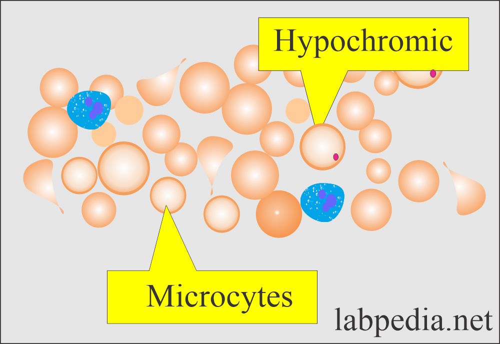 Microcytic hypochromic RBC |
|
|
|
|
|
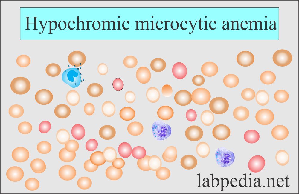 Mean corpuscular volume (MCV): Anemia hypochromic microcytic |
|
|
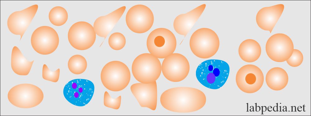
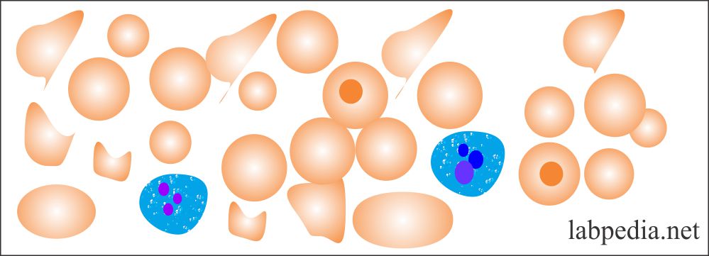 RBC poikilocytosis |
|
|
 RBC Anicytosis |
|
|
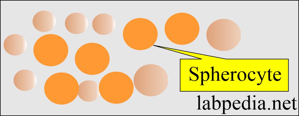 RBC spherocytes |
|
|
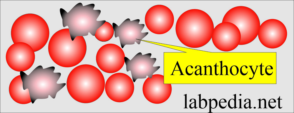 Peripheral blood smear: RBC acanthocyte cells |
|
|
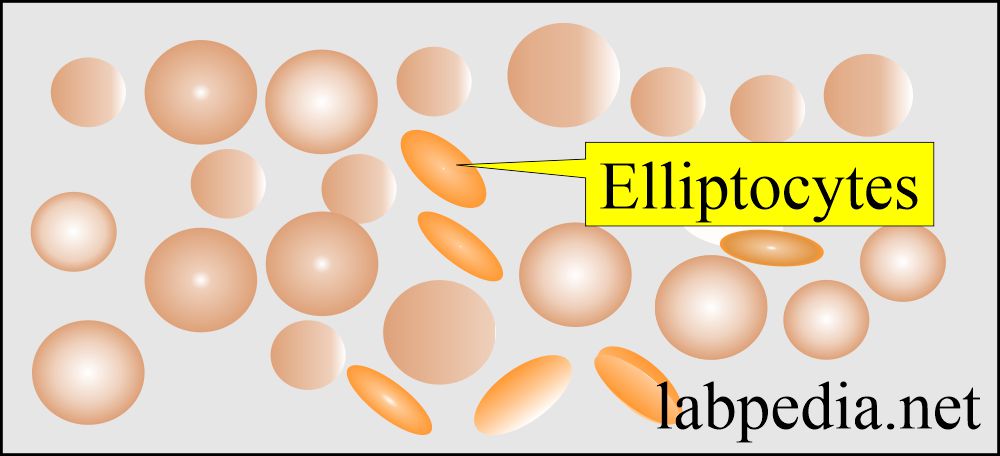 Peripheral blood smear: RBC Elliptocytes |
|
|
Nice more and more know this subject
Thanks.
Thankyou for your information
Thanks.