Parasitology:- Common Parasites Pictures
Common Parasites Pictures
What sample is needed to find common parasites?
- Preferably, take a fresh stool sample.
- Thick and thin peripheral blood smears.
- The serum for serological studies.
What are the disadvantages of stool examination?
- It will not differentiate between present and past infection.
- Can not find drug resistance.
- May not find causative agent.
What precautions are needed for stool examination?
- Foreign materials in the stool may create problems like barium, bismuth, mineral oil, and non-absorbable antidiarrheal drugs.
- The use of drugs like tetracycline may change the flora if given one week before the stool examination.
A short review of common Parasites is described.
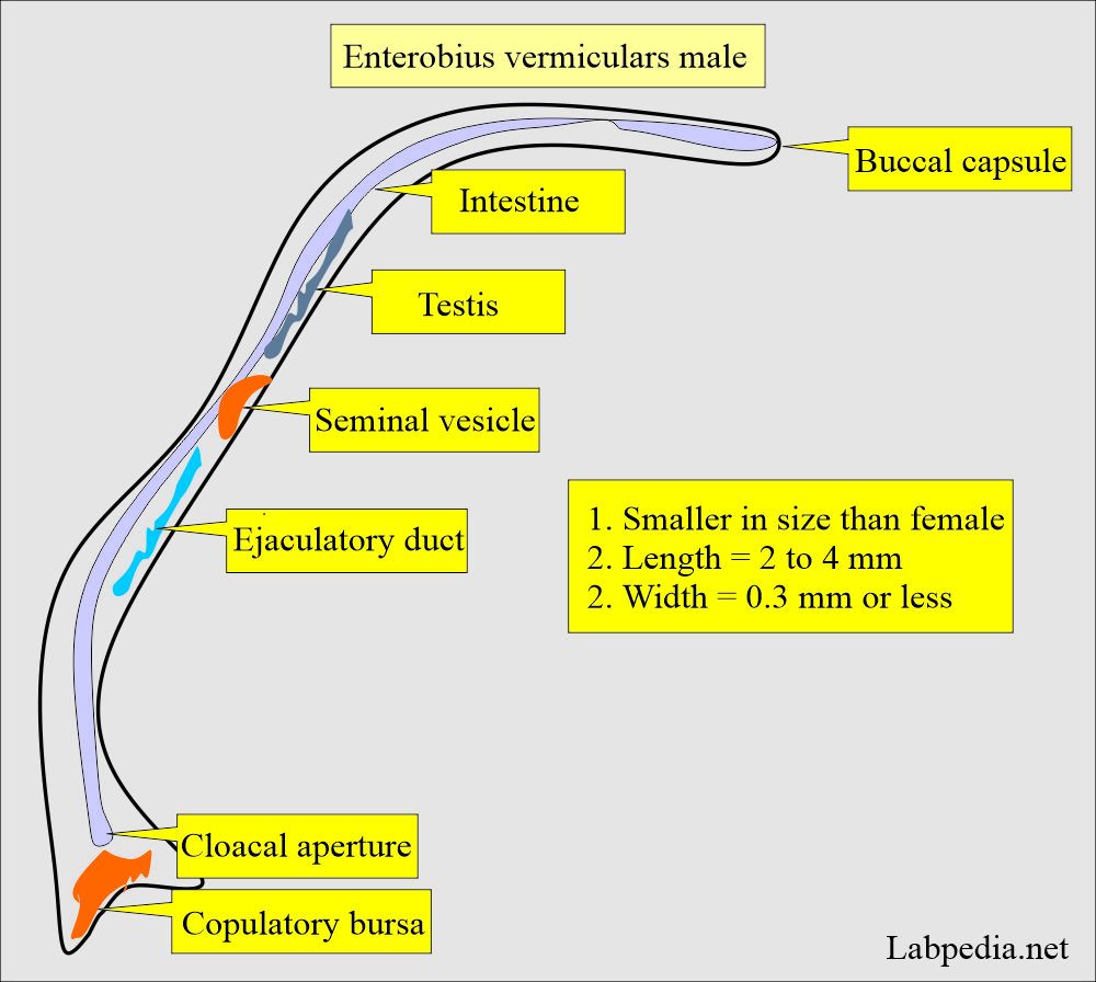
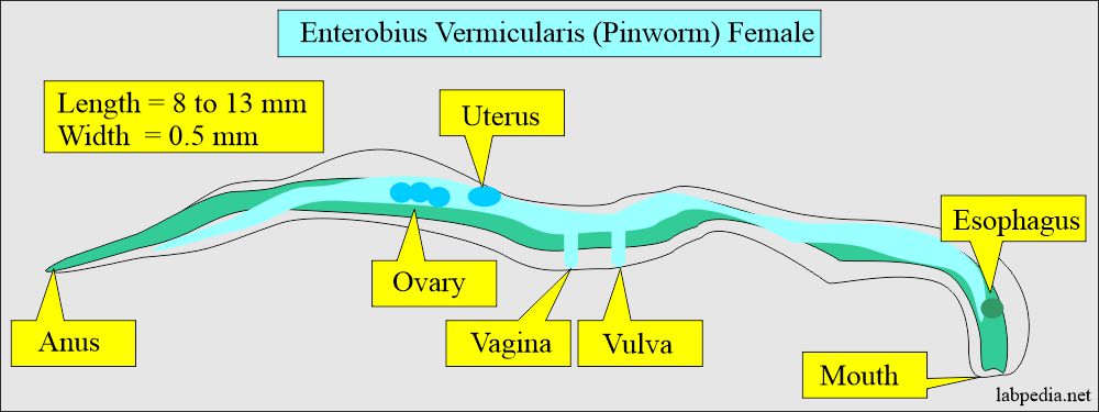
Enterobius Vermicularis
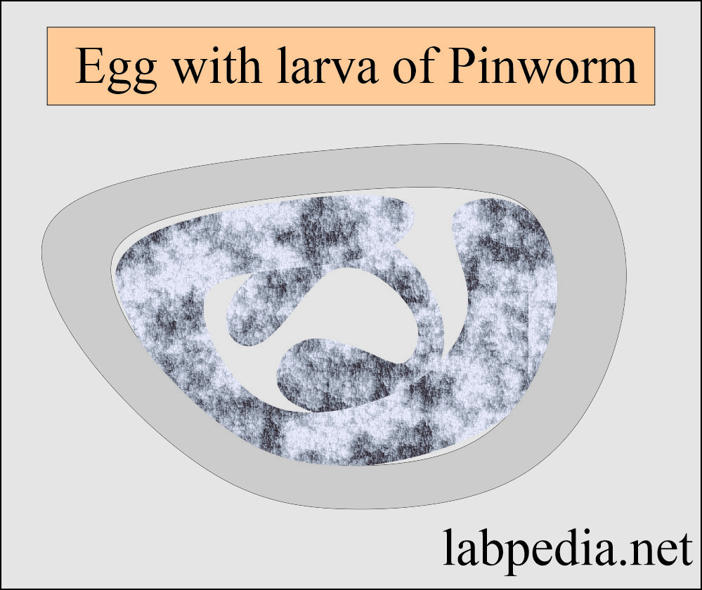
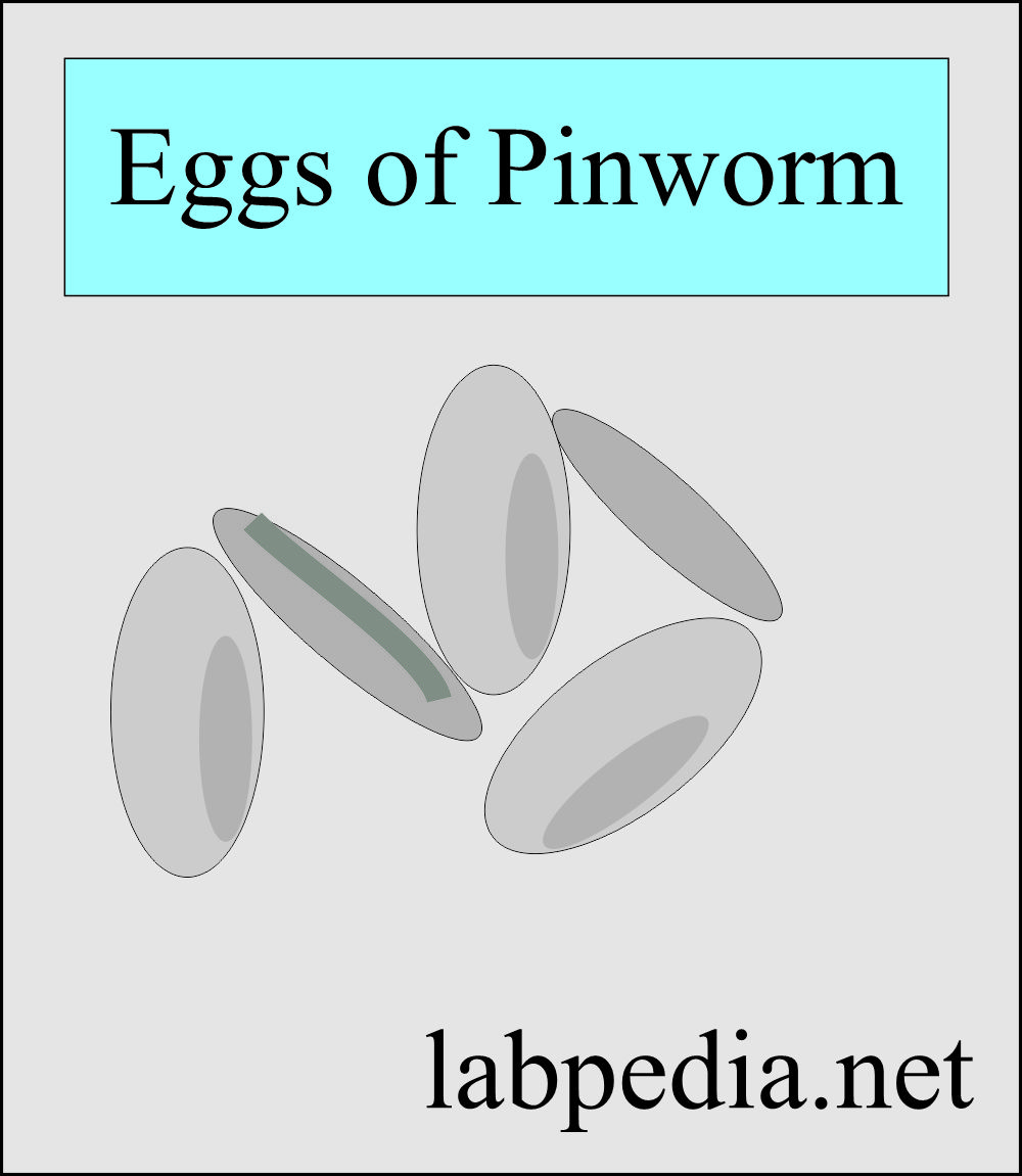
- The ova is oval and asymmetric, with one side flattened.
- Size is 55 × 26 micrometers.
- In underdeveloped countries, 10% of the pediatric population may be infected.
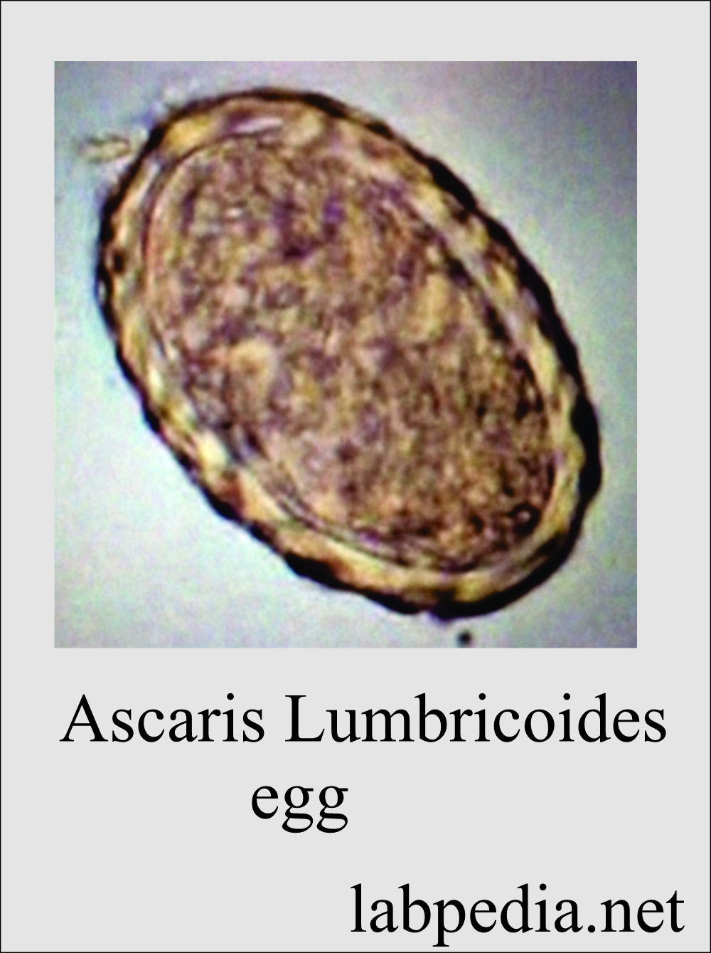
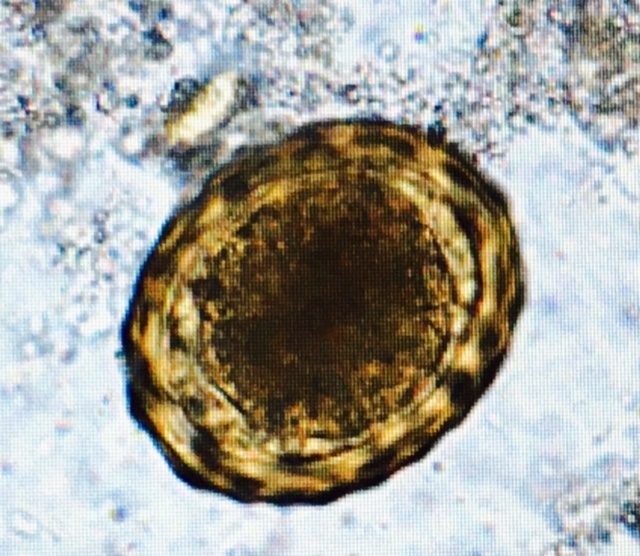
Ascaris Lumbricoides
- It is a large intestinal roundworm. It has a common resemblance to common earthworms.
- The fertile ovum is 60 x 45 micrometers. It has a thick shell and is surrounded by an albuminous covering.
- The infertile ovum is 90 x 40 micrometers. This is surrounded by an irregular albuminous covering.
- Female worm ranges from 20 to 35 cm in length.
- It may be as thick as a lead pencil.
- Males are rarely more than 30 cm in length.
- Males are more slender and have prominent incurved tails.
Ancylostoma Duodenal
- It is called Old World hookworm.
- Ankylostoma adults are greyish-white or pinkish. The head is slightly bent in relation to the rest of the body.
- Male worm measures nearly 1 cm by 0.5 mm.
- Females are longer and stouter.
- The mouth is well developed, with a pair of teeth on either side of the median line and a smaller pair in the depth of the buccal capsule.
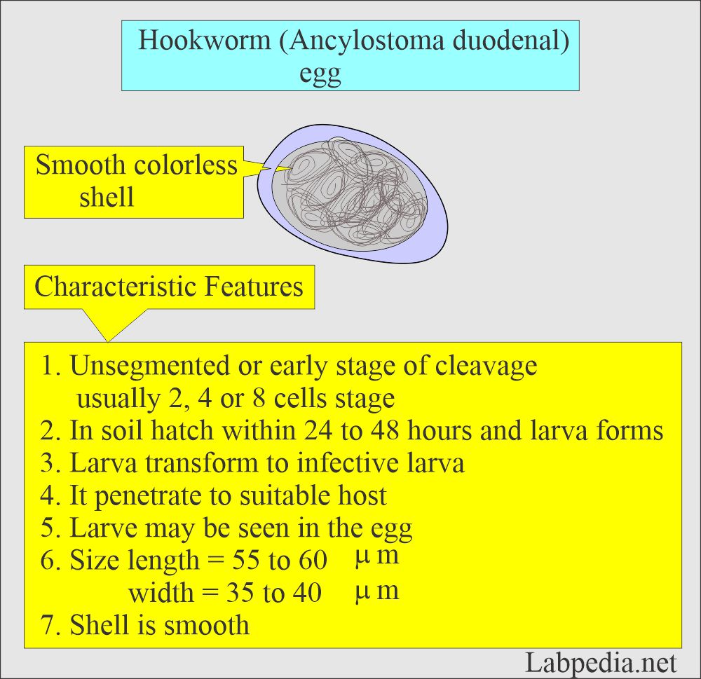
- Hookworm eggs, when passed in the stool, are unsegmented or in the early stage of cleavage.
- Ovum measures 60 to 65 x 40 micrometers. It is oval and has a thin shell.
- Usually, the embryo is at the four-cell stage.
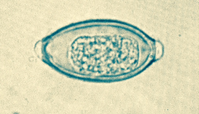
Trichuris Trichiura
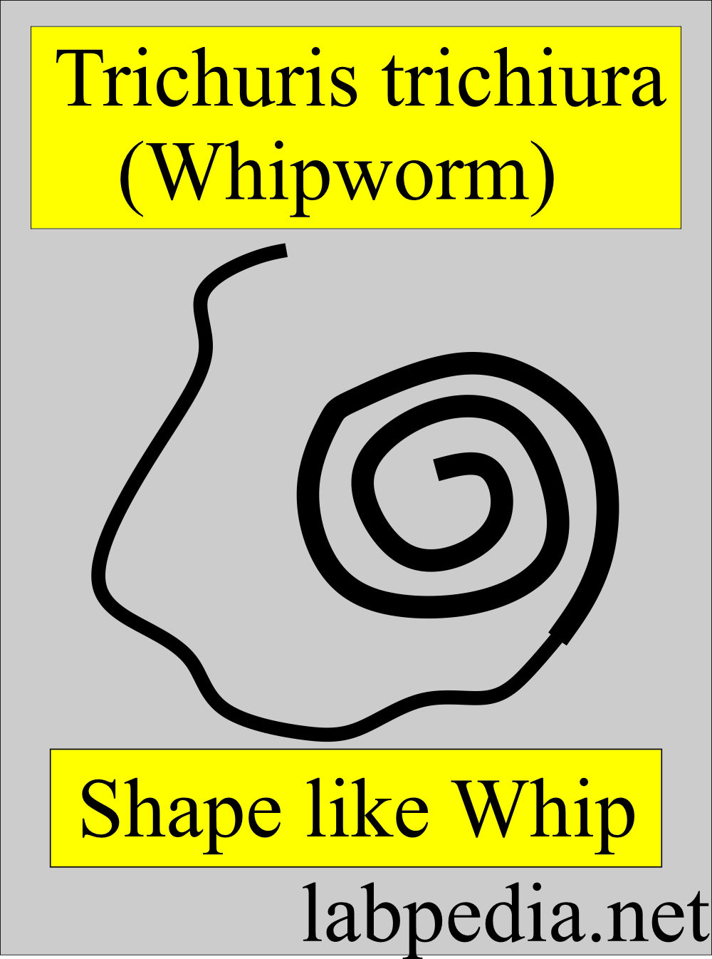
- This is also called a whipworm. It is more common in areas with poor sanitation.
- The name whipworm is very descriptive. The thick posterior part of the body forms the stock, and the long, thin anterior portion is like a lash.
- The ovum size is 52 x 23 micrometers.
- It is barrel-shaped with distinctive ends.
Giardia Lamblia
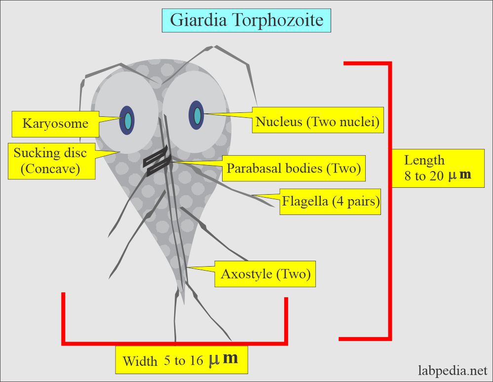
- Trophozoites are found in the upper part of the small intestine, where they live attached to the mucosa.
- These may be found in the gallbladder and biliary tract.
- Trophozoite form measures 12 to 15 micrometers. It is pyriform and has two nuclei.
- Trophozoite unstained shows progressive motility like a falling tree leaf.
- Trophozoite stained shows nuclei in the area of the sucking disc.
- There are two median bodies posterior to the disc.
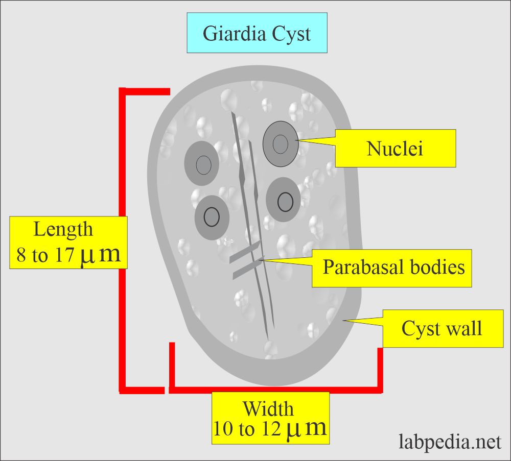
- Cysts unstained are ovoid in shape,
- Stained cysts show 4 nuclei and four median bodies.
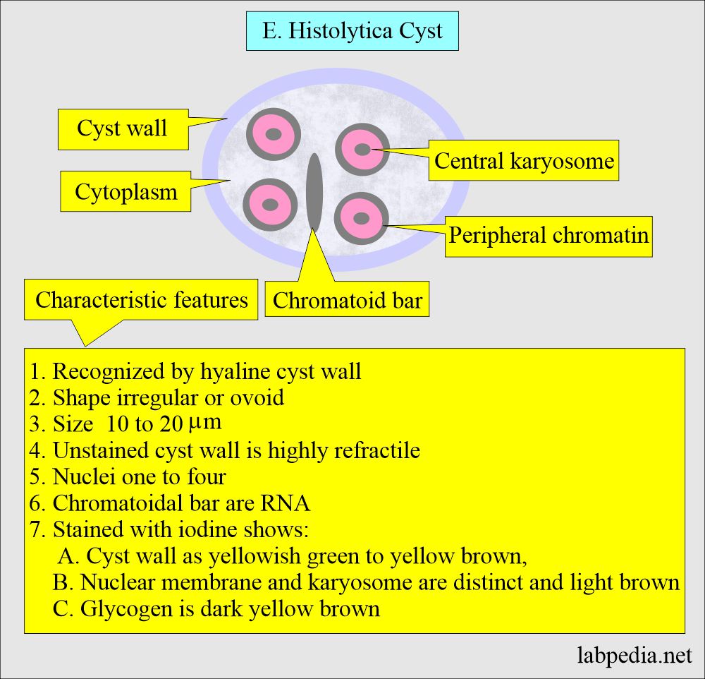
Entamoeba Histolytica
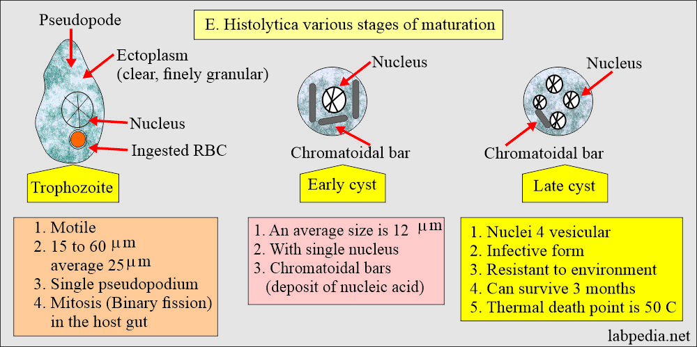
- Entamoeba Histolytica is found in roughly 10% of the world’s population—actually, it may be 1%. The rest may be due to Entamoeba dispar.
- Entamoeba Histolytica was described in 1875 in a Russian Peasant.
- Entamoeba Histolytica mainly inhabits the large intestine, where they eat RBCs and may cause ulcers.
- Entamoeba Histolytica causes chronic dysentery.
Iodamoeba butschlii
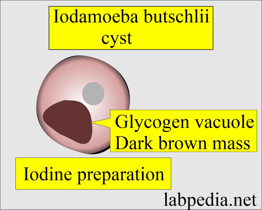
- Iodamoeba butschlii is an amoeba, and its name was given due to the presence of characteristic glycogen vacuoles in the cyst.
- Iodamoeba butschlii has variable shapes.
- It is easily recognized in the iodine preparation.
- Trophozoites in unstained smear is difficult to recognize.
- These measure 4 to 20 µm in diameter.
- These have sluggish progressive movements and have hyaline pseudopods.
- Cyst ranges from 6 to 16 µm in diameter.
- A refractile cyst wall surrounds an unstained cyst.
- These cysts are irregular in outline.
- The glycogen vacuole is prominent even in unstained cysts.
- When stained with iodine, the glycogen vacuole is a dark brown mass.
Trichomonas vaginalis
- Trichomonas vaginalis is closely related to Trichomonas Hominis.
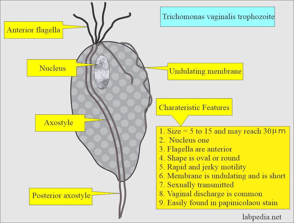
- Trichomonas vaginalis size is 5 to 15 µm but may reach 30 µm.
- In vaginal secretions, you may see jerky movements, nondirectional motility.
- It is motile due to the presence of anterior flagella.
- The movement is rapid and jerky.
Questions and answers:
Question 1: Why is Trichuris trichiura called whipworm?
Question 2: What is characteristic of Iodamoeba butschlii?

