Mycobacterium Tuberculosis:- Part 1 – Pulmonary Tuberculosis (TB)
Mycobacterium Tuberculosis (TB)
What sample is needed for the diagnosis of Tuberculosis?
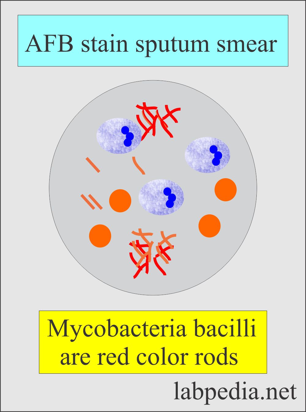
- For pulmonary tuberculosis, you can advise sputum for AFB stain.
- X-RAY chest may also help.
- A lymph node biopsy may be done to see granulomatous inflammation.
- You may see granuloma in the bone biopsy.
- Examination of the CSF.
How would you discuss the epidemiology of M. tuberculosis?
- Tuberculosis is the world’s most spreading disease and developing drug resistance disease.
- Mycobacterium tuberculosis is the causative agent.
- It is estimated that 20% to 43% of the world’s population suffers from TB.
- In the USA, 15 million people are infected (Old statistics).
- TB occurs in :
- Poor communities suffer, and these are considered to be the diseases of poor people.
- Malnourished people.
- Homeless.
- Overcrowded community.
- Substandard housing.
What is the mode of the spread of tuberculosis?
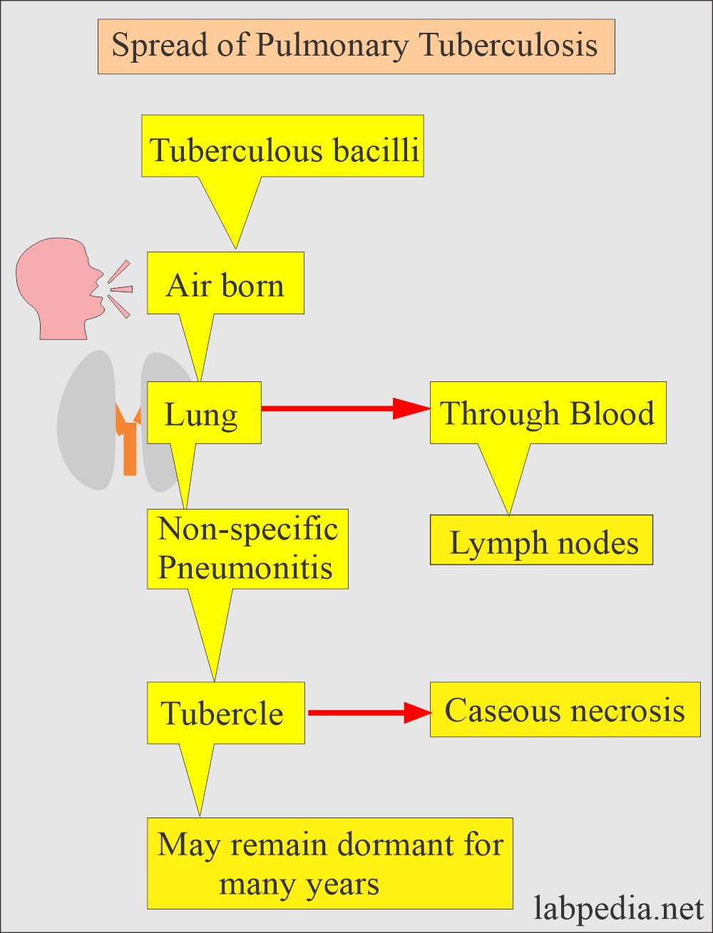
- This is an airborne disease.
- This occurs in various forms and modes.
-
- Primary TB = Clinically and radiologically, it is a silent disease.
- Latent TB = Do not have active disease and can not spread the disease to others.
- Active TB = 10% of the latent TB develop active TB when not given treatment.
- Progressive primary TB = 5% of the primary active TB with signs and symptoms.
- It is thought that 90% of the disease is a reactivation of latent TB.
What is the Microbiology of Mycobacterium Tuberculosis?
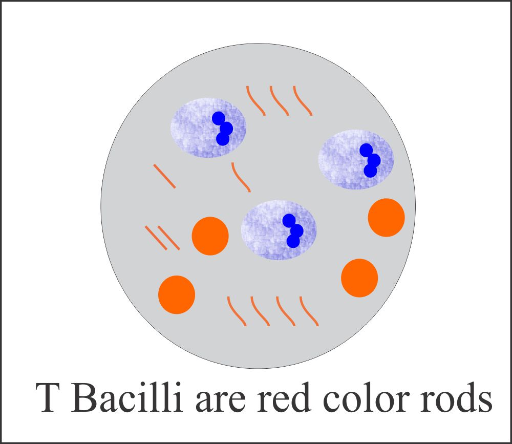
- These are acid-fast bacilli.
- These are rods shape and grow in cords.
- The growth is very slow on special media.
- They get gram stain but are very weak, which is gram stain positive.
- These are non-motile, obligatory aerobes and intracellular organisms.
- Humans are the only reservoir.
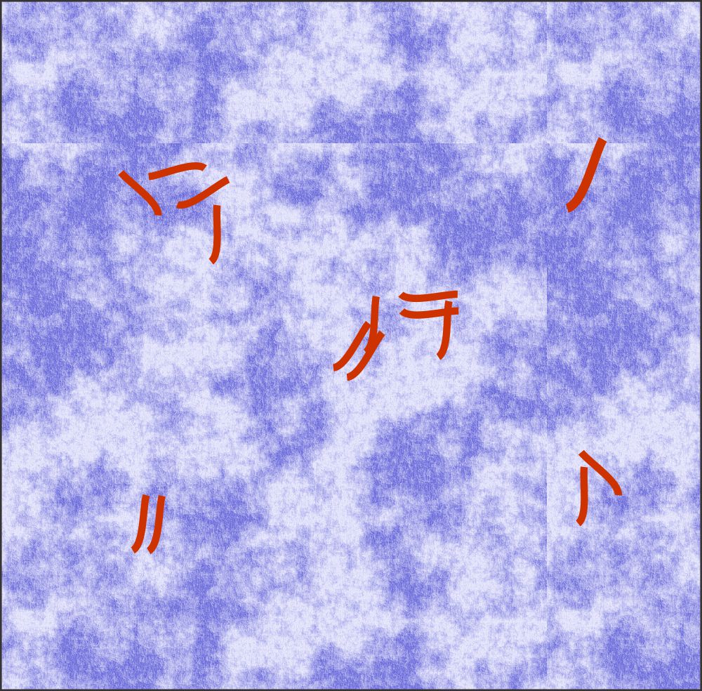
Mycobacterium tuberculosis (TB) Acid-fast bacilli
How will you classify the Mycobacteria:
Mycobacteria are divided on the basis of the disease they cause.
Slow growers:
- Mycobacterium tuberculosis causes tuberculosis in humans.
- M. Bovis leads to bovine tuberculosis.
- M. leprae leads to leprosy.
- M. avium and M. intracellularae cause disseminated infection in the AIDs patients.
- M. Kansasii leads to lung infection.
- M. marinum leads to skin infection and deeper infection, like arthritis and osteomyelitis.
Rapid growers:
- M. fortuitum and M. chelonae cause opportunistic infection and invasion of the deeper tissue.
Other classification divides Mycobacteria into:
- Mycobacteria.
- Atypical mycobacteria.
- Non-tuberculous mycobacteria.
What is the pathogenesis of Mycobacterium Tuberculosis?
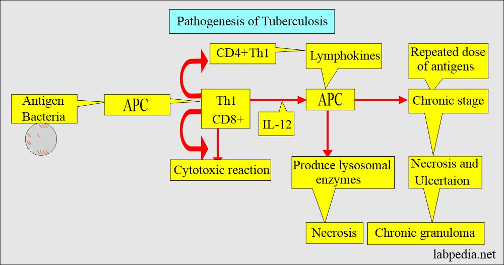
- Mycobacteria are engulfed by macrophagic cells, and in them, they survive and multiply.
- These macrophagic cells ingest the mycobacteria and carry them via lymphatics to the local hilar lymph nodes.
- In the lymph nodes, type IV hypersensitivity reaction (Cell-mediated immune response) takes place.
- Mycobacterium tubercle bacilli cause damage by invading the macrophagic cells by Type IV hypersensitivity reaction.
- This bacteria leads to caseating necrosis and granuloma formation.
- There are multinucleated giant cells, Langhans’ type cells.
- TB bacteria consists of slightly curved or straight rods.
- The gram stain cannot stain it, but it is acid-fast.
- These are nonmotile and without spores.
- Pathogenic bacteria are slow-growing and may take 4 to 6 weeks.
- The common types are :
- Mycobacterium tuberculosis.
- Mycobacterium bovis.
- others are Runyon group 1 to IV.
What are the signs and Symptoms of Mycobacterium Tuberculosis?
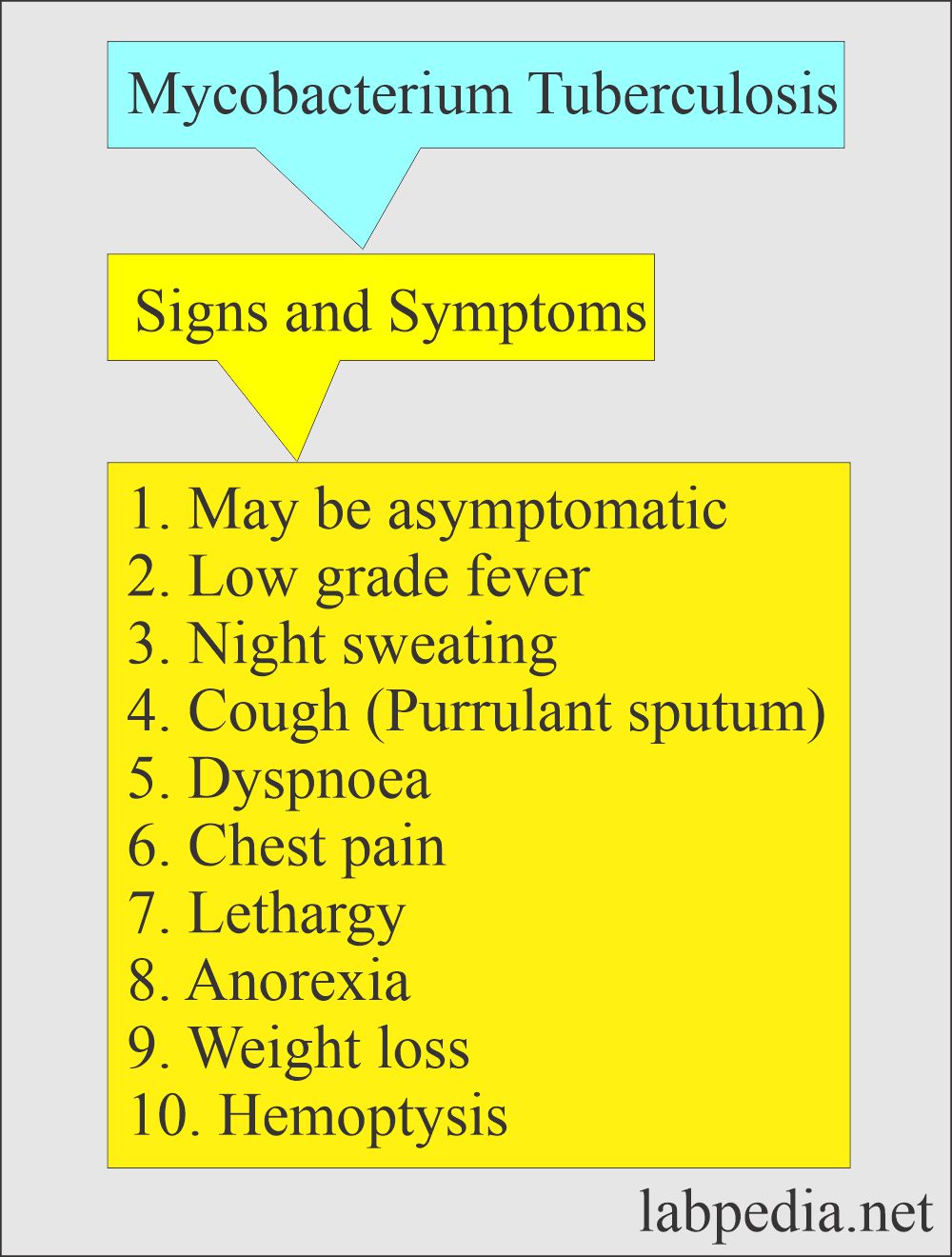
- The patient will have the following:
- Malaise.
- Anorexia.
- Weight loss.
- Fever.
- Night sweating.
- A chronic cough is a common presentation of pulmonary TB.
- Blood-streaked sputum is common.
- The patient may have hemoptysis.
- Rarely are patients asymptomatic.
- In advanced disease:
- There may be clubbing of nails.
- Enlarged lymph nodes in the neck.
- The patient may develop pleural effusion.
What are the possibilities of Mycobacterium tuberculosis infection?
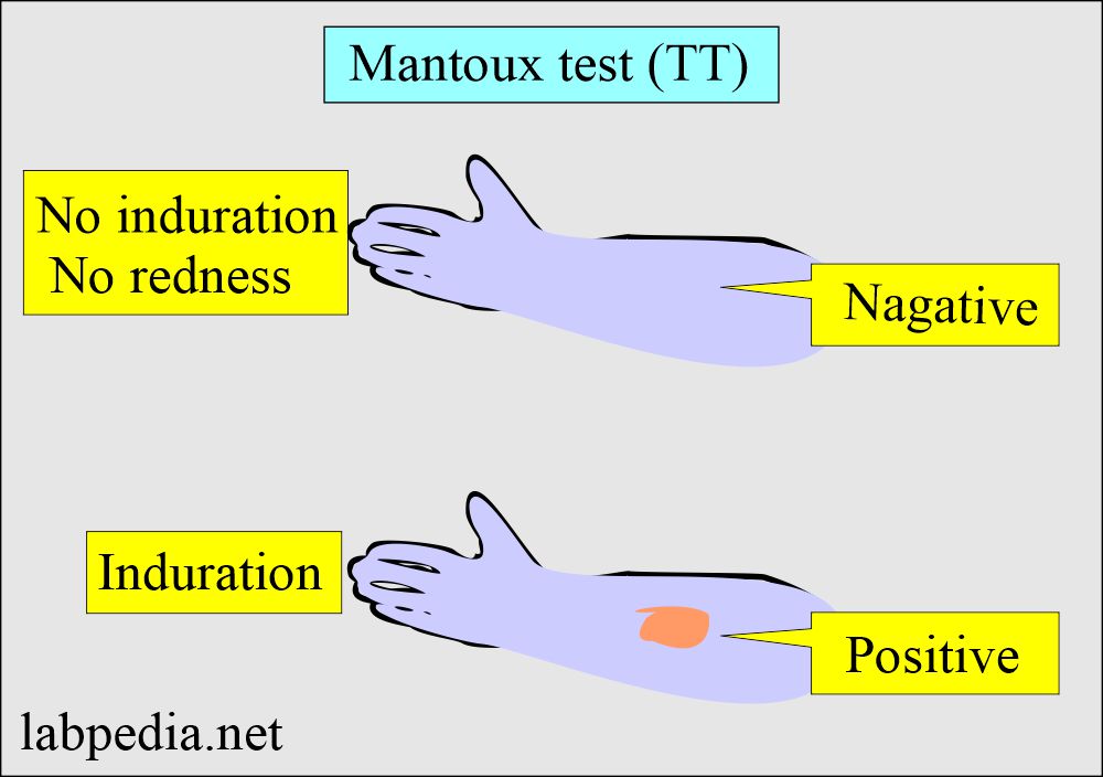
- TT (Manteaux test) positive cases, and these cases may be inactive asymptomatic people.
- Primary tuberculosis shows the Ghon complex.
- In primary tuberculosis, the disease is mostly insidious.
- There is a lesion in the lung and involvement of the lymph nodes.
- Secondary tuberculosis involves the upper lobe of the lung because of the higher oxygen concentration.
- This is usually seen in impaired immunity.
- Cavity formation may occur in the lungs.
- Sputum smears are AFB-positive.
- The disease is contagious.
- Miliary tuberculosis is a widespread disease.
- It involves the lungs, CNS, kidneys, and GI tract.
- It may involve any organs, including the bones.
- Extra-pulmonary may involve CNS and lead to chronic meningitis.
- There may be the formation of tuberculoma in the brain.
- There may be the involvement of the skin.
- TB is very common in AIDS patients.
- In AID patients, TT may be negative due to compromised immune systems.
What are the complications of tuberculosis?
- Primary tuberculosis in the lungs.
- Involvement of the kidneys.
- Involvement of the neighboring part of the lungs.
- Pleural effusion.
- Involvement of the meninges.
- Brian abscess.
- Involvement of the intestines.
- Peritonitis.
- Skin infection
How would you diagnose Mycobacterium Tuberculosis?
- Definite diagnosis depends upon the demonstration of T.Bacilli by:
- Culture.
- Culture on solid media needs 12 weeks.
- Culture on liquid media needs several days.
- PCR by DNA or RNA amplification method.
- Sputum, three consecutive samples is recommended for:
- Fluorochrome staining with rhodamine-auramine.
- AFB stain or Ziehl-Neelsen stain.
- An early morning specimen is recommended.
- Bronchoscopy is advised for bronchial washing in case of negative sputum.
- Transbronchial lung biopsy increases the diagnostic yield.
- Gastric aspiration. An early morning sample is an alternative to bronchoscopy.
- Blood culture: 15% of the cases may indicate a positive culture for T. bacilli.
- The sensitivity should be done once the culture is positive.
- The sensitivity should be assessed if the sputum culture is positive after 2 months of treatment.
- Needle biopsy of the pleura shows granulomas in 60 % of the cases.
- Pleural fluid cultures are positive in < 25 % of the cases.
- Radiology: The chest X-ray shows a small homogenous opacity.
How will you describe the significance of the Mantoux test or Tuberculin test (TT)?
- The Mantoux test (TT) will not distinguish between latent and active TB.
- 0.1 ml (5 tuberculin units) of PPD should be injected intradermally.
- The best site is the volar surface of the arm.
- Injected with 27 G needle.
- Read after 48 to 72 hours for induration (thickening of the injected area).
- After the infection, it takes 2 to 10 weeks to develop an immune response to PPD.
- Other specimens that can also be used are:
- Urine. The first-morning clean catch is collected for three consecutive days.
- Stool. This should be collected in a clean, sterile container.
- Blood. Lysed centrifuged blood is used for culture.
- Niacin test. Mycobacterium produces Niacin. Commercially available kits can test this.
What are the preventive measures for Mycobacterium Tuberculosis?
- Health workers can prevent this disease.
- If your TB test is positive, you have contact with the patient and may not have active disease.
- Vaccination like BCG is helpful in preventing the disease.
- BCG is live attenuated bacteria.
- It is not available in the USA.
How would you treat Mycobacterium Tuberculosis infection?
- The following drugs are used in patients with tuberculosis:
- Isoniazid (INH).
- This is advised in TT-positive people.
- This is also given prophylactically in AIDs patients.
- Combination of drugs like:
- Isoniazid.
- Rifampicin.
- Pyrazinamide.
- Ethambutol.
- Isolation is also important to stop the spread of the disease.
- Steroids are contraindicated in these patients because there may be reactivation of tuberculosis.
- Isoniazid (INH).
What is the outcome of tuberculosis?
- There is a steady decline in the incidence of tuberculosis because of the:
- Specific preventive measures.
- Improvement in social conditions.
- Use of the BCG vaccines in the prevalent area.
- Use of isoniazid for 1 year with a close contact person.
Questions and answers:
Question 1: How much time for the culture of tubercle bacilli?
Question 2: When to read Mantoux test (TT)?

