Lyme Disease Diagnosis
Lyme Disease Diagnosis
What sample is needed for Lyme Disease Diagnosis?
- Whole blood or citrated blood is needed.
- Other samples are CSF and synovial fluids in the acute stage.
What are the precautions for Lyme Disease Diagnosis?
- Spirochetal diseases like syphilis or leptospirosis can give a false-positive reaction.
How will you define Lyme disease?
- Lyme disease is caused by spirochete Borrelia burgdorferi by several tick vectors.
- The most common vector deer in the Northeast and North-central United States of America is the deer tick, Ixodes dammini.
- The Pacific coast states have Ixodes pacificus, the western black-legged tick, morphologically called hard tick.
What is the causative Agent of Lyme Disease?
- Lyme disease was first diagnosed in Lyme, Connecticut, in 1975.
- The spirochete Borrelia burgdorferi causes Lyme disease.
- The bite of a deer tick causes this disease (Ixodes dammini or pacificus).
- These ticks are the best vector for Lyme disease.
- The bite of a deer tick causes this disease (Ixodes dammini or pacificus).
- This isn’t easy to grow in the culture.
- It takes a long time to grow.
- Cultural success is in 50% of the cases.
- The culture of blood and CSF has a poor result.
- Serological tests are more helpful.
- Its diagnosis was established in early 1980.
How will you discuss the epidemiology of Lyme disease?
- The three major affected areas are:
- Northeastern states like New Jersey to Connecticut.
- Far Eastern states.
- Upper midwestern states.
- Cases are reported in Canada, Europe, and Australia.
What are the reservoirs of Lyme Disease?
- Rodents and deer serve as the natural reservoir for these ticks. Other animals are sheep and cattle.
- In the Northeast and North-central United States of America, the deer tick is called Ixodes dammini.
- In the Pacific coastal states, it is called Ixodes pacificus.
- It is also reported in Canada, Europe, and Australia.
- Ixodes dammini has a 2-year cycle with three forms of life.
- The very young tick is called the larval stage.
- Dormant stage until the following spring.
- The larval stage changes into the Nymph stage, which has 4 pairs of legs like an adult stage.
What is the morphology of Borrelia (Lyme Disease)?
- These are 0.18 to 0.25 X 4.3 μm. These are flexible helical spirochaete.
- These are gram-negative organisms.
- Culture: These are microaerophilic, growing at 34 °C in a special medium.
- Ixodes dammini has 2 years, three-form life cycle.
- Larval stage: The very young tick is called the larval stage.
- The larval ticks are very small and have only 3 pairs of legs like insects.
- Nymph stage: These ticks have four pairs of legs in the following years, like in the adult stage.
- Adult stage: These ticks are 4 legged.
What is the presentation of Lyme disease?
- In 50% to 80% of the patients, after one week of the bite by ticks (range 3 to 68 days), a reddish macular spreading lesion with central clearing appears (erythema chronicum migrans).
- This chronic inflammatory disease first shows a distinct skin lesion, Erythema migrans (erythema chronicum migrans).
- It starts at the site of the bite as a red macule.
- There is a central clearing at the site of the bite.
- This lesion usually fades within 2 to 3 weeks and is usually accompanied by low-grade fever, weakness, fatigue, and regional lymphadenopathy.
- This characteristic skin lesion should strongly suggest Lyme disease.
- 10% of patients develop anicteric hepatitis.
- 7% of the patients develop transitory ECG abnormality or myocardial inflammation after about 5 weeks of the tick bites.
- In the second stage of illness, aseptic meningitis or peripheral nervous system abnormalities like Bell’s palsy or Bannwarth’s polyneuritis syndrome occur after about four weeks of tick bites.
- Erythema migrans.
- Late Lyme disease.
- Lyme arthritis: There is migratory arthralgia, and myalgia is frequently present.
- Cardiovascular involvement.
- Neurological involvement and presentation.
- In the third stage of the disease, about 40% of the patients develop recurrent arthritis.
- This is the most common involvement of one or more joints, with the knee being the most common site.
- The joint involvement starts after 6 weeks to 6 months after the tick bites.
What are the signs and symptoms of Lyme Disease?
- This disease was first time diagnosed in Lyme, Connecticut, in 1975.
- Skin lesion: This disease usually starts in summer with skin lesions called Erythema chronicum migrans.
- This lesion usually appears at the site of the deer tick bite.
- Characteristically, there are:
- Fever.
- A headache and a stiff neck.
- There is fatigue.
- There is muscle and joint pain.
- These patients may develop arthritis and meningitis.
- Migratory arthralgias and myalgias are frequently seen in these patients.
- About 10% of the patients develop anicteric hepatitis.
- There are chronic meningoencephalitis and peripheral neuritis.
- In CVS: There are myocarditis, pericarditis, and conduction defects.
- About 7% of the patients develop ECG changes or myocardial inflammation, usually after 5 weeks of the tick bite.
- Arthritis stage: In the third stage of the disease, about 40% of the patients develop recurrent arthritis.
- This is the most famous sign that arthritis of large joints is present, and it is often recurrent.
How will you diagnose Lyme Disease?
- ESR is raised in 50% of the cases.
- WBCs are raised in about 10% of the cases.
- Aspiration of the joint synovial fluid has findings similar to those of rheumatoid arthritis.
- CSF:
- When there are meningeal or peripheral nerves, symptoms usually show;
- Increased WBCs predominantly lymphocytes.
- Normal glucose.
- Mildly increased protein.
- An oligoclonal band like multiple sclerosis.
- CSF IgM antibodies are present.
- The culture:
- Blood culture is positive in the second stage of the disease.
- CSF culture is positive in the second stage of the disease, with S/S in about 10% of the cases.
- Biopsy of the erythema migrans skin lesion can be used for culture.
- The transport medium used is, in particular, BSK.
- The culture medium used in Kelly’s medium and BSK II.
- Antigen detection: Urine shows excreted B. burgdorferi.
- PCR: Polymerase chain reaction (PCR). It shows 80% positivity when the sample is taken from the skin lesion.
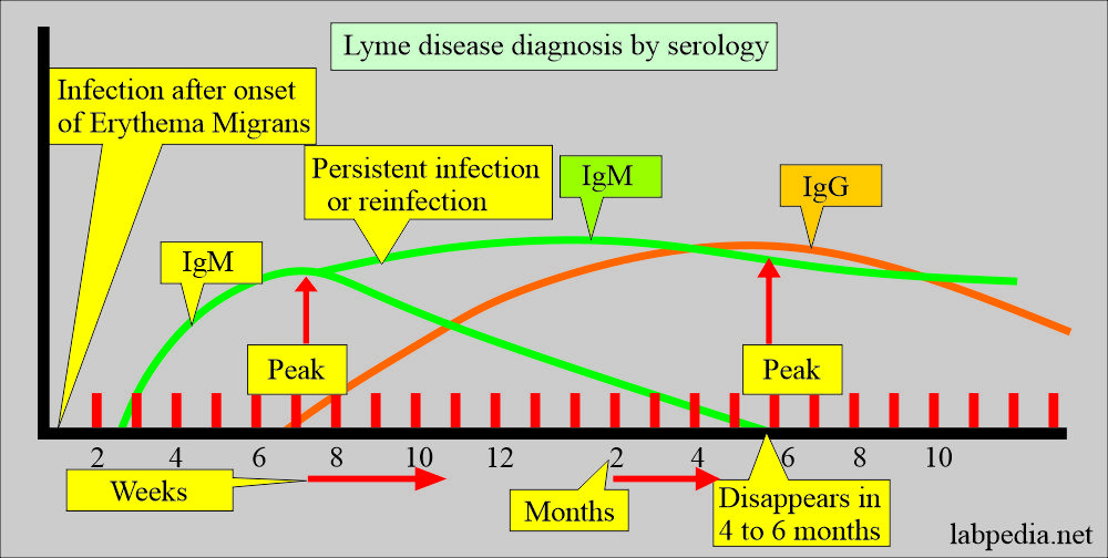
- Antibody detection: This is the most common method.
- It is negative when there is a low level of IgM and IgG antibodies.
- IgM titer peak is from the 3 to 5th week of disease onset.
- Then, it declines.
- A single high titer of specific IgM is diagnostic.
- IgG is low during the first few weeks of the disease and reaches the maximum level by 4 to 6 months later on.
- In the early stage, the serologic tests are rarely positive.
- Indirect immunoassay.
- Enzyme immune assay has replaced the indirect immunoassay. ELIZA is the preferred method of diagnosis.
- The western Blot method is the confirmatory test.
- Skin biopsy of the affected area surrounding the erythema migrans.
- Silver stain reveals spirochetes.
- The special stain used is Warthin-Starry silver stains for a skin biopsy.
- CDC criteria:
- Isolation of B. burgdorferi.
- The Positive IgM and IgG antibody in Blood or CSF.
- The positive antibody titer is in the convalescent or acute stage sera.
- Test results positive are:
- In the earliest stages of the disease, in the case of skin lesions, positivity is rare.
- After the onset of skin lesion, 3 to 4 weeks after onset is around 40%.
- In the second stage of the disease with systemic disease, around 65% are positive.
- In the third stage of the disease, it is around 90% to 95%.
- One of the studies shows the following results:
- Active Lyme disease = 23%.
- Previous Lyme disease = 20%.
- No evidence of Lyme disease = 57%.
How will you treat Lyme Disease?
- Mostly, this disease is treated with amoxicillin and doxycycline for 3 weeks.
- Some time may need IV therapy.
- These are also sensitive to penicillin and tetracycline.
Questions and answers:
Question 1: What are findings of Lyme disease in case of arthritis in the synovial fluid?
Question 2: What is the difference between the nymph stage and the larval stage of the tick?


