Gram Stain
What sample is needed for Gram Stain?
- Gram stain can be done on sputum, pus, tissue, and urine.
- The sample can be obtained from the infected ulcer or wound.
- The CSF may be stained.
What are the indications for Gram stain?
- Gram stain differentiates between gram-positive and gram-negative organisms.
- To diagnose the presence of bacteria in sputum, pus, or any other tissue or fluids.
- To diagnose bacterial meningitis.
- It can stain yeast, and this needs to be reported.
What is the history of Gram stain?
- Bacteria are colorless and usually invisible to light microscopy, so a staining color was developed to show those bacteria.
- The name comes from its inventor, Hans Christian Gram. He published a gram stain method in 1884.
- He was searching for the organism and was diagnosed with pneumonia.
- This is a special stain for diagnosing gram-positive or gram-negative organisms in various samples, such as sputum, pus, urine, etc.
- The bacteria that absorb the crystal violet and hold on to it will appear blue, called gram-positive organisms.
- If the crystal violet is washed off with alcohol, it will absorb the counterstain safranin, a gram-negative organism.
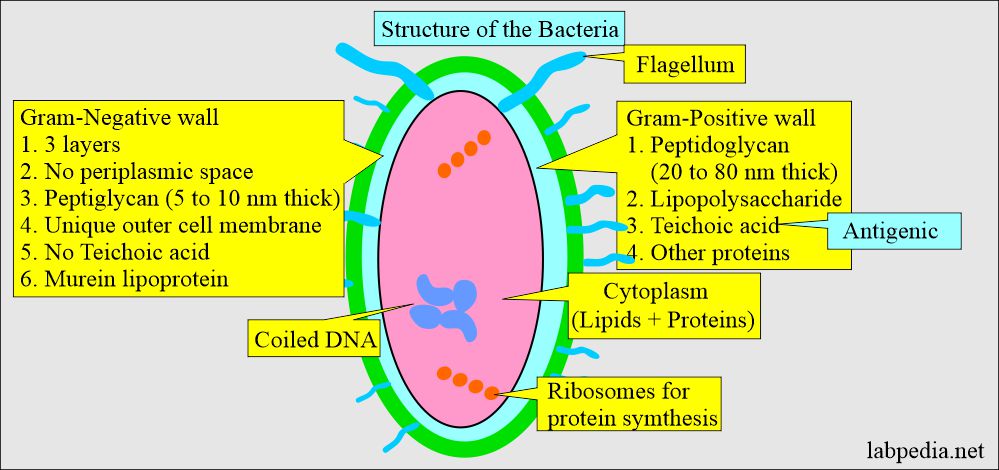
What is the structure of the bacteria?
- Bacteria are prokaryotes (single-cell organisms).
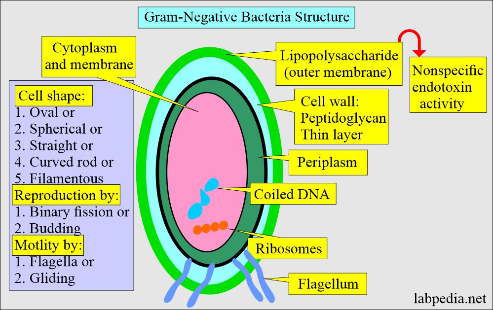
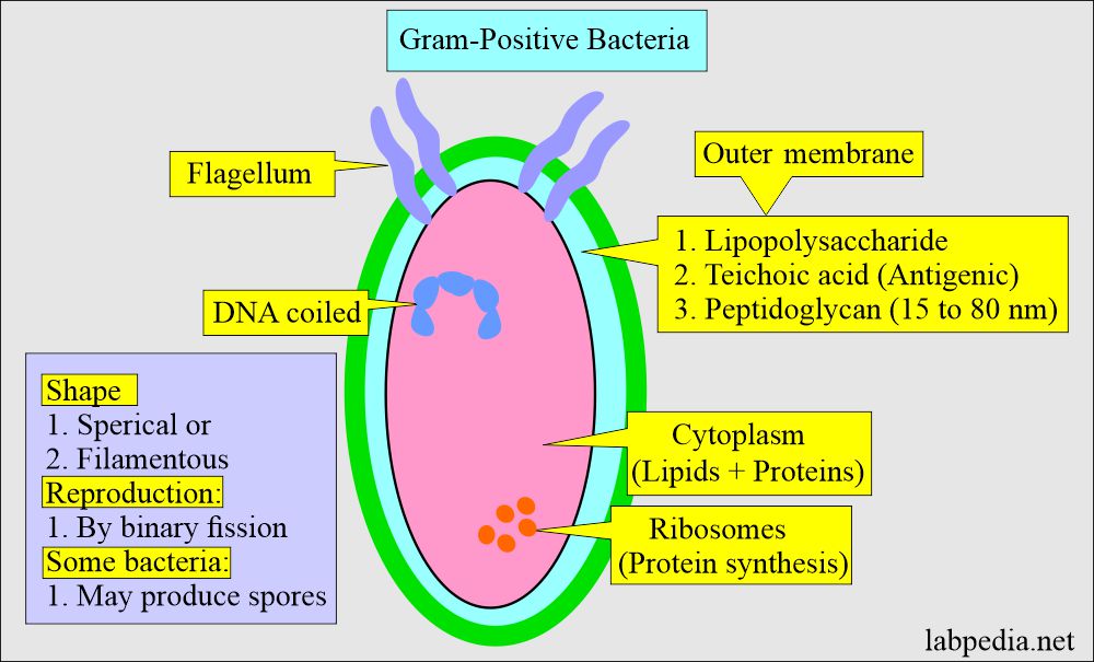
- The layer outside the cytoplasm is called the peptidoglycan layer and is present in gram-positive and gram-negative bacteria.
- Gram-positive bacteria have a thick wall and extensive cross-linking of the amino acid chains. In contrast, gram-negative bacteria have a very thin wall and a simple cross-linking pattern.
- Bacteria consist of circular DNA molecules (Continuous coding of the gene).
- DNA is tightly coiled.
- Only ribosomes are seen, which is needed for protein synthesis.
- Gram-positive organisms are hydrophilic, and this property prevents bacteria in the intestine from bile.
- Gram-negative bacteria’s outer cell walls are also hydrophilic, but the lipid component molecules give hydrophobic properties.
How will you classify the bacteria?
- Gram-positive = Blue.
- Gram-negative = Red.
- The gram-negative bacterial wall consists of three layers.
- Gram-positive bacteria consist of Two layers.
How will you differentiate Gram-positive and Gram-negative bacteria?
| Characteristic features | Gram-positive | Gram-negative |
|
|
|
|
|
|
|
|
|
|
|
|
|
|
|
|
|
|
|
|
|
|
|
|
|
|
|
How will you do Gram staining?
It consists of the following steps:
- Fix the slide by heat.
- Primary stain: Stain the slide smear with gentian violet or crystal violet. It will penetrate the cell membrane.
- Mordant: wash the violet stain and flood the smear with the Iodine solution. This acts as a mordant and forms a complex with Crystal violet.
- Decolorizer: Wash off the smear and flood it with alcohol (95 %) or an Acetone-alcohol mixture.
- This will remove the outer cell membrane in gram-negative bacteria, where the complex will be washed off.
- Gram-positive bacteria’s cell membrane remains intact, and the stain will not be washed off after alcohol treatment.
- Decolorization with acetone or alcohol will lead to the following:
- Gram-positive bacteria block the dye extraction (still, this step is unclear).
- This step will decolorize gram-negative bacteria and not gram-positive bacteria.
- Secondary stain: Counterstain the smear with safranin O, a red dye.
- The counterstain cannot enter gram-positive bacteria, so the bacteria are purple.
- In gram-negative bacteria, safranin can enter and give a pink color.
How will you report the results of the gram stain?
- Gram-positive bacteria are blue-purple.
- Gram-negative bacteria are pink magenta.
How will you give some examples of gram-stain bacteria?
| Type of microorganism | Morphology in gram stain |
|
|
|
|
|
|
|
|
|
|
|
|
|
|
|
|
|
|
|
|
|
|
How will classify bacteria based on gram stain?
| Bacteria | Gram-positive | Gram-negative |
|
|
|
|
|
|
What are the examples of Bacterial Infections?
- Gram-positive cocci infections:
- Staphylococcal aureus can cause skin infections.
- Toxic shock syndrome.
- Gram-negative cocci infections:
- N.meningitidis causes meningitis.
- N.gonorrhoeae causes gonorrhea.
- Gram-negative bacilli infections:
- E.coli causes urinary tract infection.
- Gram-positive bacilli infections:
- B.anthracis leads to skin infection and pneumonia.
- Listeria monocytogenes may cause food-borne infection.
- Viruses do not stain with gram stain.
Questions and answers:
Question 1: Who was the inventor of the gram stain?
Question 2: What is the role of safranin counter staining for gram-negative bacteria?


