Fluid Analysis:- Part 5 – Amniotic fluid Examination (Amniocentesis)
Amniotic fluid Examination (Amniocentesis)
What sample is needed for Amniotic fluid Examination (Amniocentesis)?
- Amniotic fluid is collected by amniocentesis.
- Amniotic fluid needs to be refrigerated.
- Avoid amniotic fluid from light for the estimation of bilirubin.
- Amniocentesis is done during the 14th, 16th, and 18th weeks of gestation.
- Chorionic villus sampling is better than amniotic fluid for karyotyping and genetic analysis.
What are the Indications for Amniotic fluid Examination (Amniocentesis)?
- To diagnose the genetic disorder (cytogenetics analysis).
- To diagnose chromosomal abnormalities.
- To diagnose inherited metabolic disorders like cystic fibrosis.
- To diagnose neural tube defects like myelomeningocele, anencephaly, and spina bifida,
- Measure bilirubin in Rh sensitization for erythroblastosis fetalis.
- To diagnose chorioamnionitis.
- To assess fetal lung maturity, a sample was taken after 32 weeks. The lecithin/sphingomyelin ratio is a measure of fetal lung maturity. Phospholipids are measured in amniotic fluids.
- To assess postmature pregnancy >40 weeks.
- To find intrauterine retardation.
- To find congenital infections like:
- Toxoplasmosis.
- Cytomegalovirus (CMV).
- It can do a culture for bacterial infections.
- Amniocentesis helps with elective abortion in a defective fetus.
- To diagnose respiratory distress syndrome (RDS). This is a complication in premature newborns.
How will you define amniotic fluid?
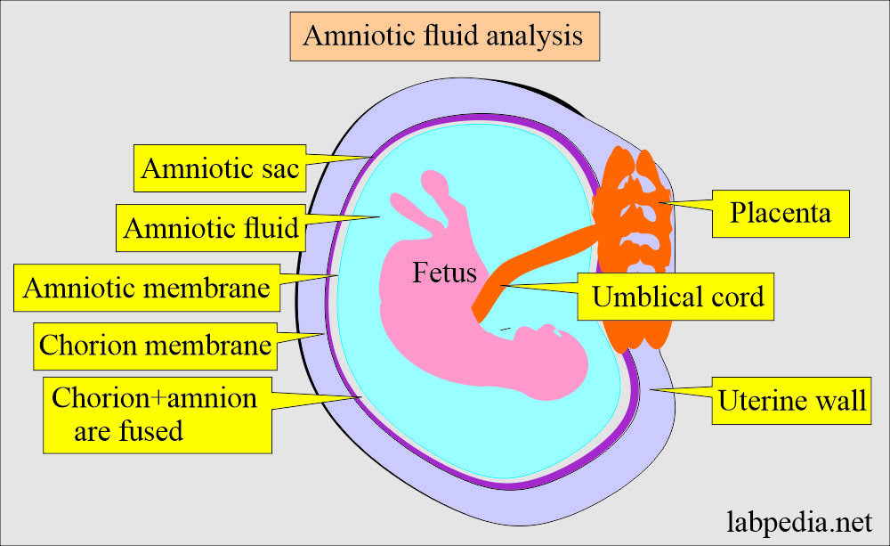
- Amniotic fluid is the fluid in the amniotic sac that surrounds the fetus.
- A portion of the amniotic fluid arises from the fetal respiratory system, urine, the amniotic membrane, and the umbilical cord.
- Amniotic fluid at term is 500 to 1100 mL.
How will you discuss the pathophysiology of Amniotic fluid?
- Amniotic fluid is present in the amnion, a membranous sac surrounding the fetus.
- Formation of amniotic fluid:
- Amniotic fluid arises from:
- Fetal respiratory tract.
- Urine.
- From amniotic membrane.
- From umbilical cord.
- Amniotic fluid arises from:
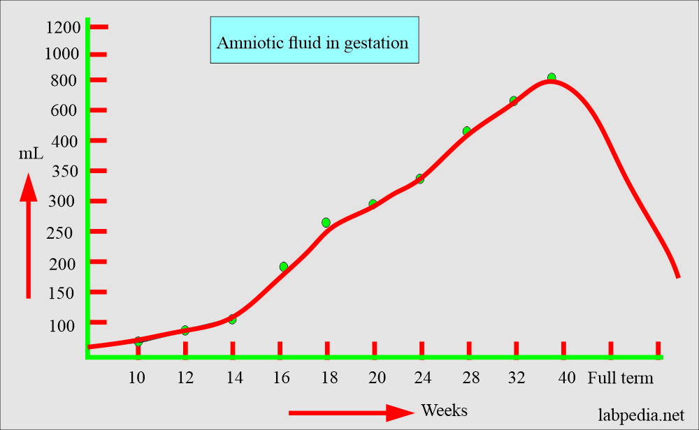
- Amniotic fluid keeps increasing throughout the pregnancy and reaches a peak of around one liter (1 L) during the third trimester.
- It gradually decreases before delivery.
- Around 30 mL of the amniotic fluid is derived from the maternal circulation during the first trimester. Its composition is similar to the mother plasma with the addition of the fetus cells. These cells may provide the basis for cytogenetics analysis.
- After the first trimester, fetal urine is the major contributor to the amniotic fluid volume.
- Now, fetal swallowing of amniotic fluid begins, and it regulates the increase in the fluid from the fetal urine.
- Hydramnios:
- If there is a failure in swallowing amniotic fluid, it will accumulate the amniotic fluid around the fetus called Hydramnios.
- This will lead to fetal distress, which is usually seen in the neural tube defect.
- It will occur in 1% of the pregnancy.
- This may occur in diabetic mothers.
- Oligohydramnios:
- It is decreased amniotic fluid seen in increased swallowing of fluid, urinary tract abnormalities, and membrane leakage.
- This may occur due to the abnormality of maternal, fetal, or placental complications.
How will you describe Amniotic fluid composition?
- In early pregnancy, amniotic fluid is produced by the amniotic membrane covering the placenta and umbilical cord.
- It changes when urine formation starts.
- With the advance of pregnancy, amniotic fluid is a by-product of fetal pulmonary secretion, urine, and metabolic products from the intestinal tract.
- initially, amniotic fluid is produced by the amniotic membrane and later by the maternal blood.
- Initially, volume from 30 mL increases to 350 mL by the 20th week of gestation.
- After 20 weeks, the amniotic fluid volume varies from 500 mL to 1000 mL.
- Creatinine, urea, and uric acid concentrations increase while glucose and protein concentrations decrease.
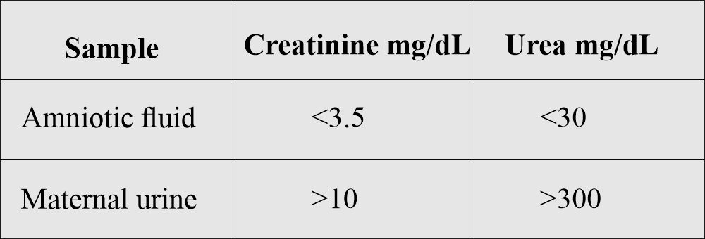
- The concentration of urea and creatinine is much lower in the amniotic fluid than in the maternal urine.
- Amniotic fluid contains albumin, urea, uric acid, creatinine, lecithin, sphingomyelin, bilirubin, fats, fructose, epithelial cells, leucocytic enzyme, and lanugo hairs.
What are the functions of amniotic fluid?
- Amniotic fluid surrounds, protects, and nourishes the fetus during gestation.
- A part of the amniotic fluid derived from:
- The fetal respiratory system.
- Amniotic membrane.
- Urine.
- Umbilical cord.
- A fetus can move freely in the amniotic fluid of the uterus.
- Amniotic fluid provides cushions to the fetus from trauma and injury.
- This will prevent the compression of the umbilical cord.
- Amniotic fluid maintains the temperature as well.
- Amniotic fluid provides information about the health and maturity of the fetus.
What is the significance of Amniotic fluid analysis?
- It can diagnose erythroblastosis fetalis, fetal maturity, and genetic counseling.
- It also diagnoses so many other abnormalities.
What are the important facts about Amniotic fluid?
- Amniotic fluid contains protein, hormones, nutrients, and antibodies.
- There is water transfer between intrauterine compartments:
- The placenta.
- The fetus.
- Amniotic fluid.
- As pregnancy (gestation) advances, then there are changes as follows:
- After 25 weeks, there is an increase in amylase, alkaline phosphatase, urea, uric acid, creatinine, and phospholipids.
- There is a decrease in chloride, bilirubin, protein, glucose, and sodium.
- An increased AFP level suggests neural tube defects, while a decreased AFP level is associated with increased risk for trisomy 21.
- Amniocentesis helps with elective abortion in a defective fetus.
- The lecithin/sphingomyelin (L/S) ratio:
- It measures fetal lung maturity.
- Lecithin is the major surfactant required for alveolar ventilation. In the case of deficiency of surfactant, alveoli collapse during expiration and lead to respiratory distress syndrome(RSD).
- There is an increased concentration of lecithin between 32 weeks to full-term babies, and there is a slight decrease in the sphingomyelin during the same period.
- Sphingomyelin is not a surfactant and has no role in lung maturity.
- This may be a major cause of death in immature babies. As the L/S ratio decreases, the risk of RDS increases.
- This test is very cumbersome.
- An L/S ratio value of 2.0 indicates lung maturity.
- L/S ratio of <2.0, newborns may not develop RDS.
- Phosphatidylglycerol (PG):
- This is the minor lung surfactant of about 10%. This is entirely synthesized by the mature lung alveolar cells, which is a good indicator of lung maturity.
- Bilirubin level:
- Analysis of the amniotic fluid for bilirubin level (or absorbance at 450) in the Rh-negative mothers in later weeks of the gestation gives the severity of anemia due to Rh-incompatibility.
- Lilly plotted the graph to show the severity of hemolytic anemia.
How will you do the procedure for amniotic fluid collection?
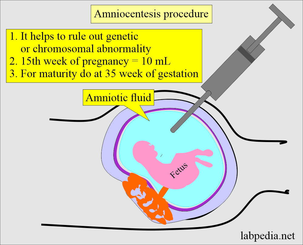
- The needle from the amniotic sac aspirates amniotic fluid is called amniocentesis.
- This is transabdominal procedure (amniocentesis).
- Another route is transvaginal amniocentesis. This method carries a great risk of infection.
- This procedure is safe if performed after the 14th week of gestation.
- Fluid for chromosomal analysis is collected after the 16th week of gestation.
- In case of fetal distress and maturity are collected in the 3rd trimester.
- 30 mL of amniotic fluid is collected in the sterile syringes.
- The first 2 to 3 ml collected is discarded because this may be contaminated by the maternal blood, tissue fluid, and cells.
- In the case of hemolytic disease of the newborn, the sample should be avoided from light. The sample should be kept in an amber-colored bottle or test tube.
- The sample for fetal lung maturity should be placed on ice for delivery to the laboratory and kept in the fridge.
- The sample for the cytogenetics may be maintained at room temperature (37 °C) before analysis.
- The sample for the biochemical testing should be separated immediately from the cellular elements and debris to avoid the effect of cellular metabolism.
What are the Complications of the amniocentesis procedure?
- There may be a miscarriage.
- There are chances for injury to the fetus.
- There may be a leak of amniotic fluid.
- There are chances for abortion.
- There are chances for premature labor.
- There are chances for infections.
- There may be amniotic fluid embolism.
- There are chances for maternal Rh isoimmunization.
- There may be damage to the urinary bladder or intestine.
What are the precautions and contraindications of amniocentesis?
- Fetal blood contamination can cause a falsely raised level of AFP.
- Do not perform this test in case of:
- Placenta previa.
- Patients with placenta abruptio.
- Patients with an incompetent cervix.
- Patient with a history of premature labor.
What are the normal values of the amniotic fluid?
The mean volume of amniotic fluid during gestation:
| Weeks of gestation | Quantity of fluid |
|
|
|
|
|
|
|
|
|
|
|
|
|
|
|
|
Source 2
- 15 weeks of gestation = 450 mL of amniotic fluid
- 25 weeks of gestation = 750 mL of amniotic fluid
- 30 to 35 weeks of gestation= 1500 mL of amniotic fluid
- Full-term = <1500 mL of amniotic fluid
Normal Amniotic Fluid findings:
| General test | Result |
|---|---|
|
|
|
|
|
|
|
|
|
|
|
|
|
|
|
|
Abnormal amniotic fluid findings:
| Lab findings | Findings | Clinical significance |
|
|
|
|
|
|
|
|
|
|
|
|
|
|
|
|
|
|
|
|
|
|
|
|
|
|
|
|
|
|
How will you interpret the amniotic fluid analysis?
- Hemolytic anemia shows increased bilirubin.
- Rh immunization also shows increased bilirubin.
- Raised AFP shows a neural tube defect. Acetylcholinesterase can confirm the diagnosis.
- AFP may also be raised in Sacrococcygeal teratoma.
- Fetal distress: The presence of meconium gives a greenish color to amniotic fluid, indicating fetal distress.
- Can diagnose Immature fetal lung.
- Can diagnose Hereditary metabolic disorders like cystic fibrosis, Tay-Sachs disease, and galactosemia.
- Can diagnose Sex-linked disorders like hemophilia.
- Genetic abnormalities like sickle cell anemia, thalassemia, and Down’s syndrome are diagnosed.
- Polyhydramnios (>2000mL) seen in diabetic patients increases the chances of congenital abnormality.
- Oligohydramnios when the amniotic fluid is less than 300 mL at 25 weeks of gestation. This is associated with fetal renal disease.
- Genetic disorder identified by the amniotic fluid analysis.
Chromosomal anomalies diagnosis is:
|
|
|
|
|
|
|
|
|
|
Questions and answers:
Question 1: What is the main function of amniocentesis?
Question 2: What is the amount of amniotic fluid at 28 weeks of gestation?

