Enterobius Vermicularis (Pinworms), Diagnosis, and Treatment
Enterobius Vermicularis (Pinworms)
How will you take a sample for Enterobius Vermicularis?
- A fresh stool is preferred.
- Cellophane tape in the anal area for children and then transfer the material to the slide.
- The perianal area is the best place to get a sample.
- Get multiple samples to rule out pinworm infection.
- The stool sample was screened to find ova or adult females.
What is the epidemiology of Enterobius Vermicularis?
- This is a member of the Oxyurida called pinworms.
- This cosmopolitan parasitic infestation is more prevalent in temperate climate regions.
- These are common in orphanages and mental hospitals where the spread is easy.
- This is the disease of school children.
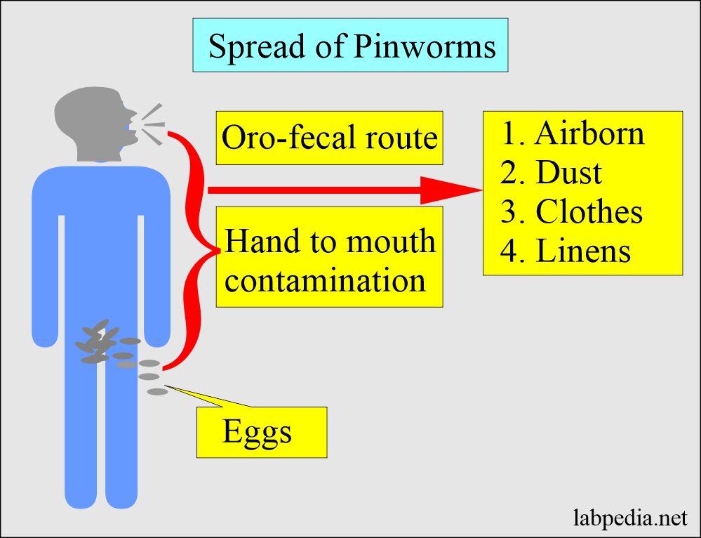
- The route of spread is oro-fecal through contaminated foods or fomites.
- Suppose inhaled then, followed by ingestion of airborne ova. Larva hatch and migrate back into the intestine.
- This is very common in the USA.
What is the morphology of Enterobius Vermicularis (Pinworms)?
- Enterobius vermicularis is also called pinworm, seatworm infection, or oxyuriasis.
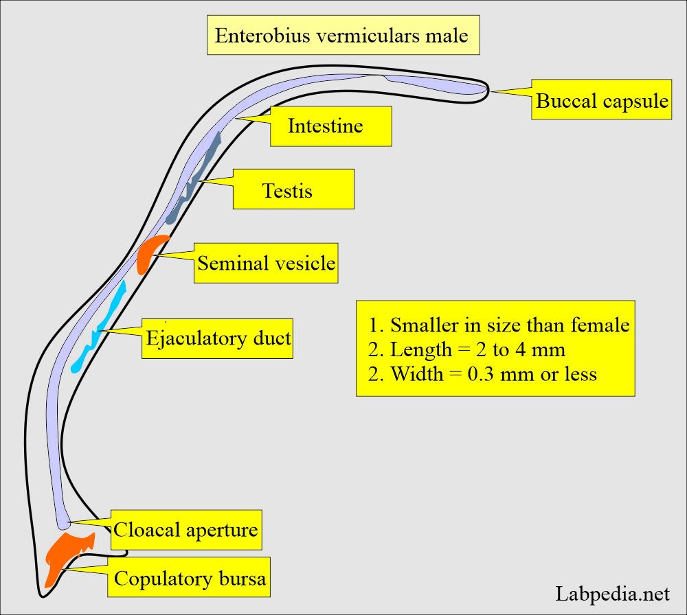
- The male measures 1 to 4 mm and has the posterior end curved ventrally.
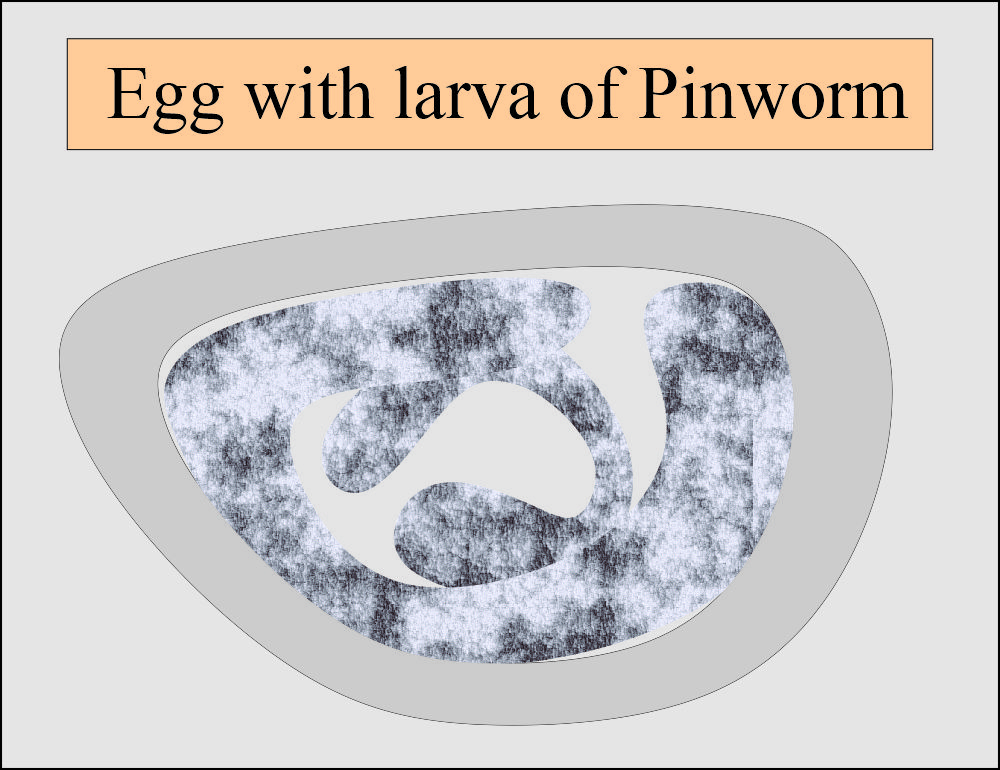
What is the measurement of the Egg of Enterobius vermicularis?
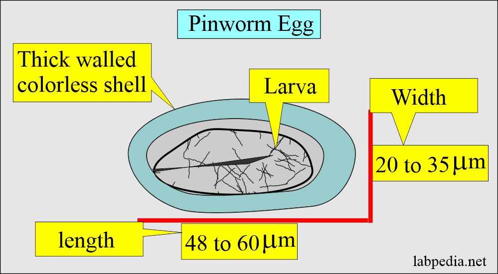
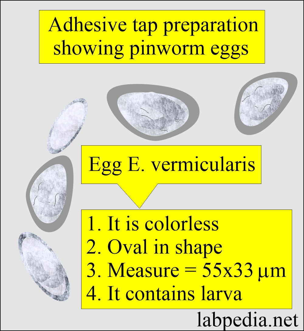
- Eggs are oval and flattened on the side.
- Length = 48 to 60 µm.
- Width = 20 to 35 µm
- The egg contains the developing larva surrounded by a thick-walled colorless shell.
- It contains various stages of the larva, unembryonated, and embryonated eggs.
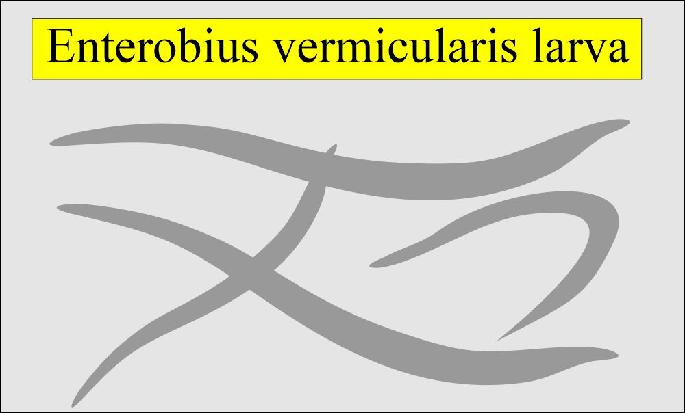
How would you describe Female Entobius vermicularis (larva stage)?
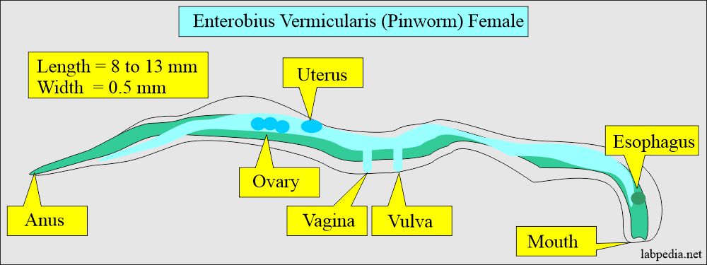
- Length = 8 to 13 mm.
- Width = Up to 0.5 mm.
- The posterior end is extended into a long, slender, pointed end, which gives it the name of a pinworm.
- The female has a vagina, vulva, ovary, uterus, and oviduct.
- There is a digestive and intestinal tract.
- There are mouth and anus.
How would you describe Male Entobius vermicularis (larva stage)?
- These are smaller in size than the female.
- Length = 2 to 4 mm.
- Width = 0.3 mm or less.
How will you explain the class Nematoda?
- The phylum is Nemathelminthes, and the class is Nematoda.
- Nematoda class has two types of parasites:
- Intestinal species:
- Enterobius vermicularis.
- Trichuris trichiura.
- Necator americanus
- Ascaris lumbricoid.
- Strongyloides stercoralis.
- Ancylostoma duodenale.
- Intestinal-tissue species:
- Dracunculus medinensis.
- Trichinella spiralis.
Discuss the Human life cycle of Enterobius vermicularis (Pinworm)?
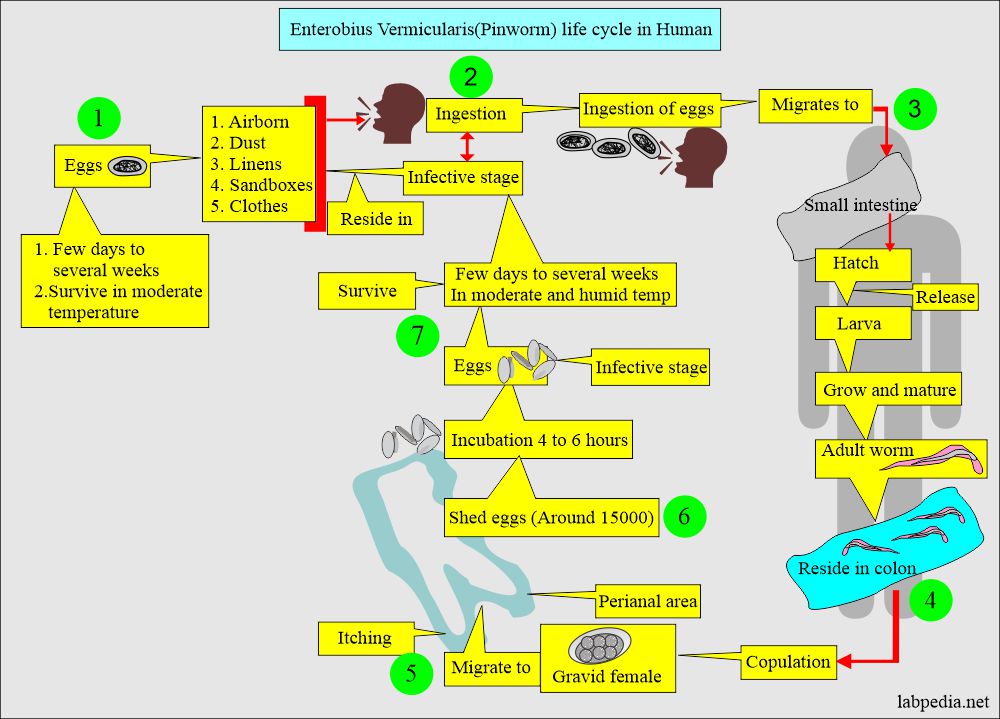
- Humans are the only hosts of Enterobius vermicularis (Pinworms).
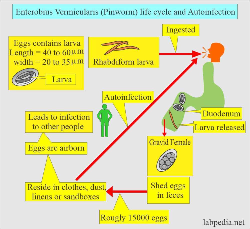
- Infective ova contains rhabdiform larva (infective eggs) ingested by humans.
- The larvae are released into the duodenum and then migrate to the lower intestine.
- These worms are attached to the intestine’s mucosa, feeding on the epithelial cells and bacteria.
- Habitat is the cecum and colon.
- The copulation of mature adults takes place in the cecum.
- The gravid female migrates to the perianal area. It sheds ova mostly at night.
- The female migrates to the anus and lays the eggs on the perianal area. There may be eggs around 15,000.
- Following incubation of 4 to 6 hours, eggs become infective.
- There may be:
- Autoinfection.
- Retroinfection
How will you explain the non-human cycle of Enterobius Vermicularis (Pinworm)?
- These ova are shed into the stool and surrounding area.
- These ova lead to infection of other people.
- The ova (eggs) are present in:
- Dust.
- Sandboxes.
- Linens.
- Clothes.
- Airborn.
What are the clinical Symptoms of Enterobius vermicularis (Pinworm)?
- Children are the most common victims of pinworms.
- It is a self-limited disease.
- Asymptomatic group:
- Some of the patients may not have any clinical symptoms.
- Symptomatic group:
- The most common symptom is perianal itching, which is very intense (pruritus ani).
- The child may have a restless sleep due to perianal itching.
- There may be pain, rashes, or skin irritation around the anal area.
- The patient may have intermittent abdominal pain and nausea.
- In the female, there may be vaginal itching.
How will you control the Enterobius vermiculars infestation?
- Personal hygiene is very important.
- Wash the hands and apply ointment to the perianal area to stop the spread of the eggs.
- Avoid scratching the infected area.
- All bedding should be washed with hot water, and cleaned the whole house to stop the spread of the disease.
- Children should wear tight-fitting diapers, pajamas, and pants to prevent contact with the perianal area. This will prevent reinfection.
- Advise washing of the anal skin area every morning soon after getting out of bed and frequently washing the clothes worn at night.
- All family members should be treated at the same time.
How will you diagnose Enterobius vermicularis (pinworm) infestation?
Procedure for adhesive tape:
- Put the cellophane tape around the anal area in cases of children. The eggs attach to the tape. Then, this tape can be seen under the microscope. Perform this test for three days.
- Or touch the slide with the tape.
- The tap will show ova 50% of the time; if you repeat this procedure thrice, positivity is 90%.
- Close the condenser iris for contrast. Otherwise, colorless eggs will not be seen.
- You can add a drop of xylene to clear the debris.
Swab method:
- Take a cotton swab, moisten it, and rub it to the perianal area.
- Wash the swab in 4 to 5 mL of saline.
- Discard the swab.
- Centrifuge the saline solution and discard the supernatant.
- Put the sediment on the slide and cover it with cover-glass
- Examine under the microscope
A stool examination:
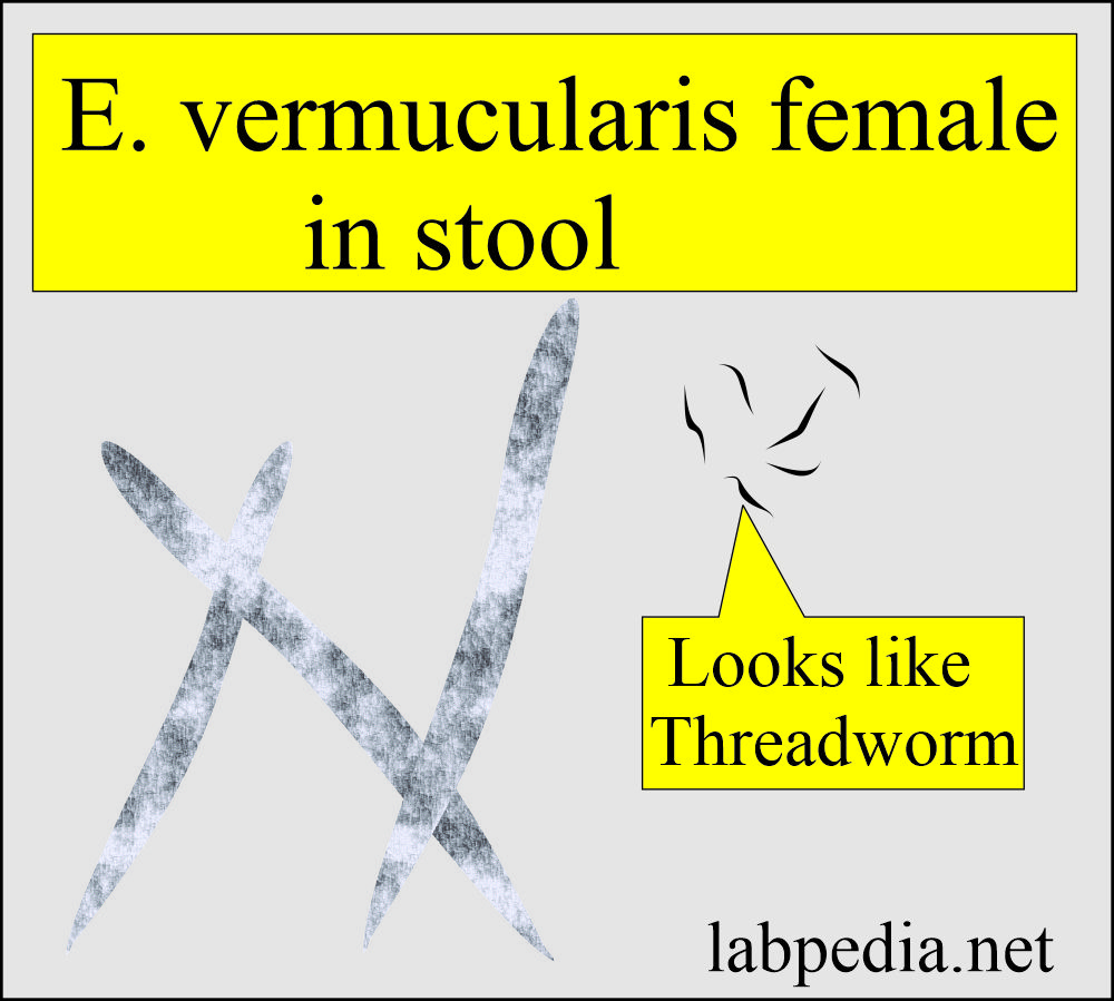
- It can show ova. May find E. vermicularis worm in the stool or while collecting the sample from the perianal area.
- These are small, measuring 8 to 13 mm in length and resembling small pieces of thread, so-called threadworms.
- These can be seen by a magnifying glass, which shows the characteristic shape, long pointed tail, and sharp anterior end.
How will you treat Enterobius Vermicularis (Pinworm)?
- The most commonly used drugs are:
- Mebendazole.
- Albendazole.
- Pyrantal pamoate.
- Piperazine.
Questions and answers:
Question 1: What is the most common presentation of pinworms in children?
Question 2: What is the outcome of the pinworm infestation?

