Cerebrospinal Fluid Analysis:- Part 2 – Cerebrospinal fluid (CSF) Normal/Abnormal Interpretations
Cerebrospinal Fluid Analysis
What sample is needed for cerebrospinal fluid (CSF) analysis?
- The sample is CSF fluid.
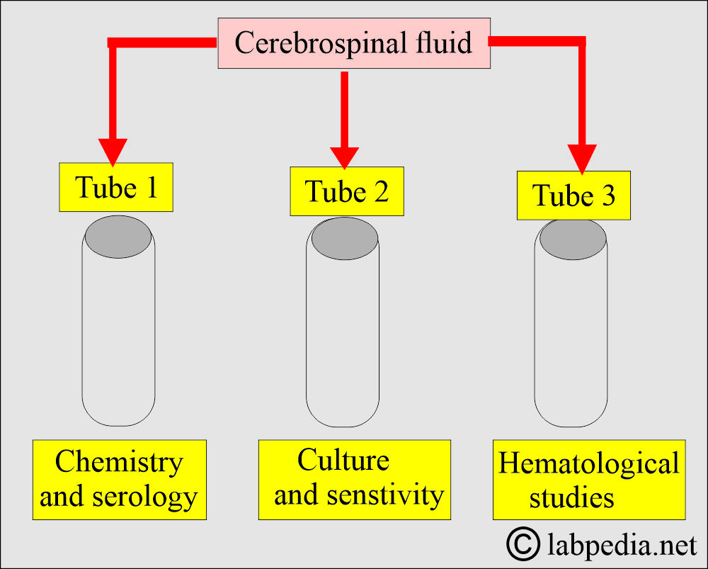
- Three tubes, each containing 2 to 3 mL of CSF, are collected. These tubes are labeled as:
- Tube 1 for chemistry and serology.
- Tube 2 for microbiology studies.
- Tube 3 for hematology studies.
- Please don’t use the first tube for culture, as it is mostly contaminated.
- The first tube can be used for chemistry and serology after centrifugation.
- The last tube is best for chemistry and microscopy because it is less likely to be hemorrhagic or contaminated.
- The third tube is ideal for hematological studies.
- Transport the sample to the laboratory as soon as possible.
Where should the CSF sample be taken from the patient?
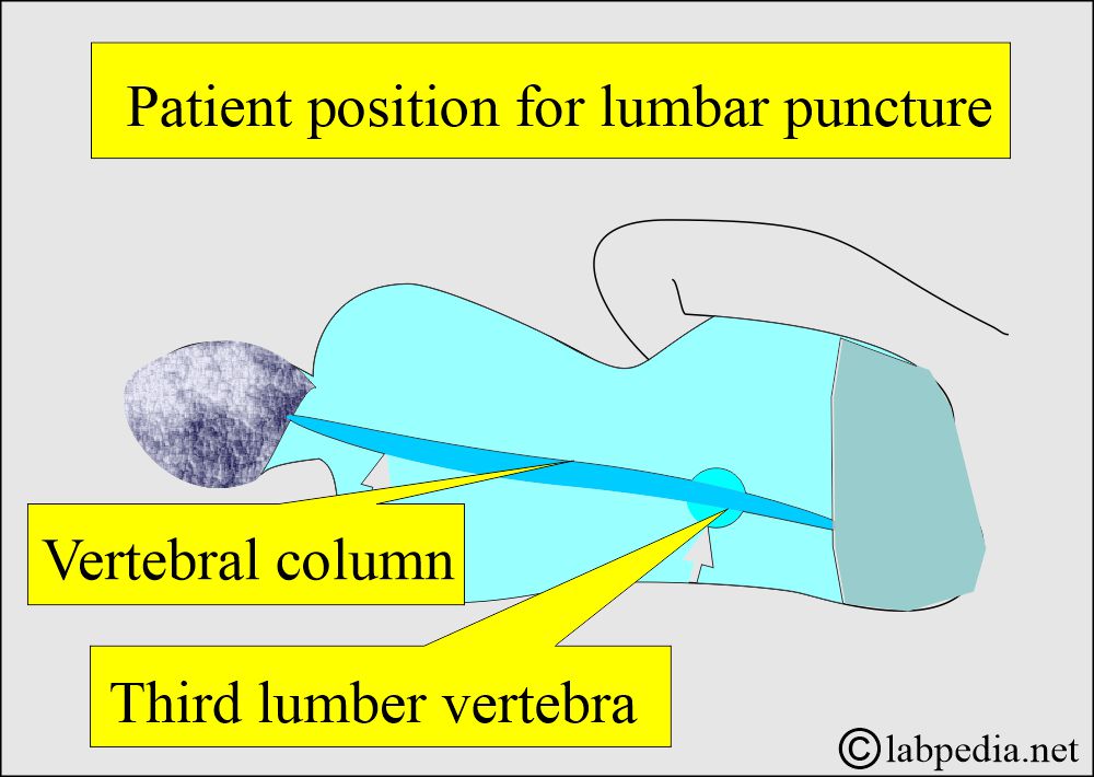
- The sample is taken from the spinal canal; the most common position is a lumbar puncture.
- After the procedure, keep the patient in a lying position in the bed for 6 to 12 hours.
What are the complications of a lumbar puncture for CSF?
- The patient may have a traumatic lumbar puncture.
- The patient may have a severe headache.
- A time may develop an infection at the site of puncture.
What are the indications for Cerebrospinal Fluid Analysis (CSF)?
- To diagnose the type of meningitis.
- To diagnose the cause of hemorrhage.
- This test is part of the patient’s workup in a coma.
- This test can diagnose cerebral malaria in infants and children.
- CSF electrophoresis is used to diagnose multiple sclerosis, particularly when an oligoclonal band is present.
What parameters are checked in Cerebrospinal Fluid Analysis (CSF)?
- CSF pressure.
- Volume.
- Appearance
- Biochemical tests include:
- Glucose.
- Protein.
- The microscopic examination gives an idea about:
- The total number of cells.
- Type of cells: Neutrophils, Lymphocytes, or RBC.
- To rule out the presence of malignant cells.
- Special stains to find bacteria (Gram stain).
- Culture.
- Special studies include:
- CSF electrophoresis for the oligoclonal band.
- Lactate dehydrogenase (LDH).
- Lactic acid.
- Chloride.
- Serology to rule out syphilis.
- Glutamine for hepatic encephalopathy in liver failure.
What are the interpretations of Cerebrospinal Fluid Analysis (CSF)?
How will you discuss the CSF pressure?
- Normal pressure is 50 to 180 mm of water.
- What are the causes of increased CSF pressure?
- Congestive heart failure.
- Obstruction of the superior vena cava.
- Cryptococcal meningitis.
- Intracranial tumors.
- Meningitis of all types.
- Cerebral edema.
- Subarachnoid hemorrhage.
- Thrombosis of venous sinuses.
- What are the causes of decreased CSF pressure?
-
- Circulatory collapse.
- Leakage of spinal fluid.
- Severe dehydration.
- Spinal subarachnoid block.
How will you check the appearance of CSF?
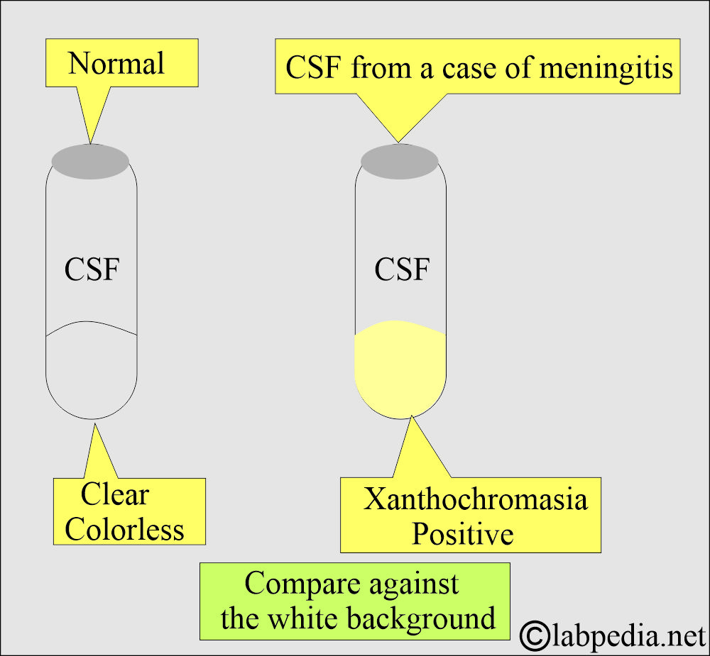
- Normal CSF is crystal clear, like water.
- The initial color of CSF is due to:
- Inflammatory diseases.
- Traumatic tap.
- Hemorrhage.
- Tumors.
How to assess the appearance of Cerebrospinal Fluid?
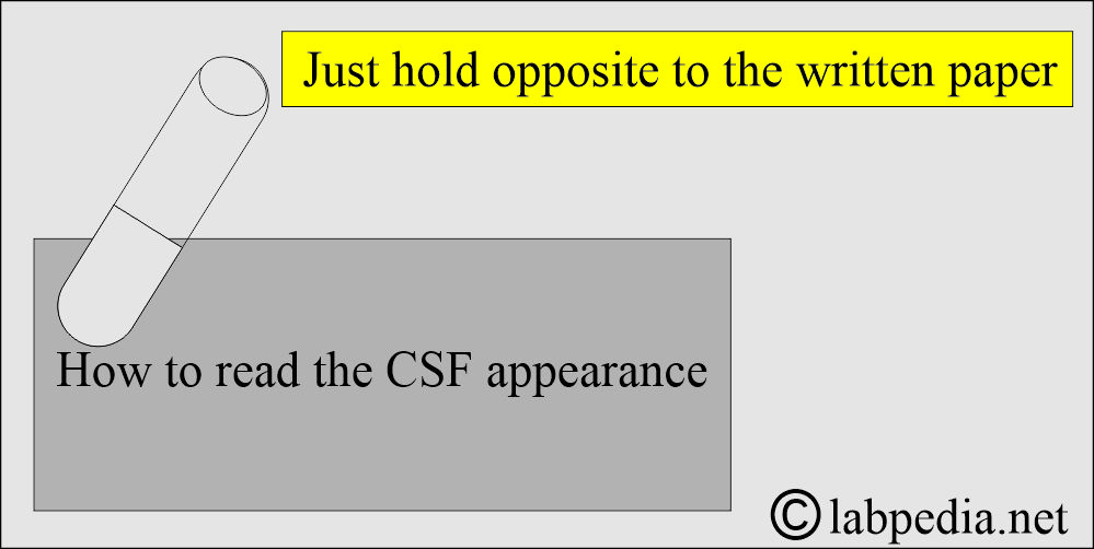
- The appearance can be compared to water.
- Hold the tube containing CSF against the paper, which can be read.
What will be the appearance of CSF in various conditions?
| Appearance | Pathological reason |
|
|
|
It may be due to the following:
|
|
|
|
|
|
|
What are the causes of various appearances of Cerebrospinal Fluid (CSF)?
- Blood-like appearance:
- Subarachnoid hemorrhage. If the sample is collected in three tubes, all the tubes will be the same color.
- Traumatic tap. Now, the third tube will be clear or less colored.
- Cloudy (Turbid) may be due to:
- The presence of WBCs.
- Increased protein.
- The presence of the microorganism.
- RBCs.
- Contrast media.
- Xanthochromia
- It is pale pink to yellow, depending on the presence of protein. This may be due to:
- Increased protein when more than 150 mg/dL.
- Bilirubin when >6 mg/dL.
- The presence of methemoglobin.
- Systemic carotenemia.
- Oxyhemoglobin due to hemolysis of RBCs.
- Melanin in meningeal melanoma.
- The yellow color may be visible in hemorrhage 10 hours to 4 weeks before the tap.
- The yellow color may also be observed if bilirubin levels exceed 10 mg/dL.
- It is pale pink to yellow, depending on the presence of protein. This may be due to:
How will you differentiate between Subarachnoid hemorrhage(SH) and Traumatic tap
- The traumatic tap may form clots, while SH does not form a clot.
- The traumatic tap is negative for xanthochromia, while SH is positive.
- An immediate repeat at a higher level will show blood in SH, whereas it is clear in the case of the Traumatic tap.
What do you know about Cerebrospinal Fluid (CSF) Glucose level?
- Glucose is utilized by bacteria (pyogenic or mycobacterium bacilli).
- Glucose may be utilized by the WBCs or occasionally by the cancer cells in CSF.
- This will take place one hour after the blood glucose.
- Glucose levels return to normal after the start of antibiotics.
- Glucose decreases by 50% in bacterial meningitis.
- CSF glucose <45 mg/dL is abnormal.
- The CSF glucose level correlates with blood glucose levels.
- This is 60% of the blood glucose.
- CSF glucose: blood glucose ratio = 0.6
- In bacterial meningitis, this ratio = <0.5
- A ratio <0.4 distinguishes acute bacterial meningitis from viral meningitis.
- Always advise blood glucose testing whenever a CSF examination is performed.
- There is a lag time between blood glucose and CSF, which is approximately one hour.
- Bacteria, more than T.bacilli, utilize glucose.
- There is no effect of viruses on the glucose level.
What is normal CSF Glucose?
- Adult = 40 to 70 mg/dL.
- Child = 60 to 80 mg/dL.
- CSF : Plasma ratio = < 0.5
- CSF glucose is less than blood glucose = 60 to 70%.
What are the causes of decreased glucose seen in CSF?
- Acute bacterial meningitis.
- Tuberculous meningitis.
- Subarachnoid hemorrhage.
- Diabetes with hypoglycemia.
- Malignant tumors with metastases to the meninges.
- Non-Bacterial meningoencephalitis.
- Syphilis.
What is the cause of Increased glucose levels?
- Diabetic hyperglycemia.
What do you know about Cerebrospinal Fluid (CSF) Protein?
- CSF protein is a nonspecific test because it is raised in so many diseases.
- CSF has a very small quantity of protein because of the blood-brain barrier.
- What are the causes of increased CSF protein?
- Increased permeability of the blood-brain barrier.
- Decreased resorption by the arachnoid villi.
- Obstruction of CSF flow.
- Increased synthesis of immunoglobulin in the intrathecal space.
- What is the normal CSF Protein?
- Adult (Lumbar area) = 15 to 45 mg/dL.
- Adult (Cisternal area) = 15 to 25 mg/dL.
- Adult (Ventricular area) = 5 to 15 mg/dL.
- Neonates (Lumbar area) = 15 to 100 mg/dL.
What are the causes of increased CSF protein?
- Traumatic tap.
- Bacterial meningitis may increase to levels of>1000 mg/dL.
- Tuberculous meningitis leads to a mild increase of 50 to 300 mg/dL.
- For fungal meningitis, the increase may be 50 to 300 mg/dL.
- Viral meningitis, the increase is mild, <200 mg/dL.
- Subarachnoid hemorrhage.
- Nonbacterial conditions like Uremia, Hypercalcemia,
- Dehydration.
- Hypercapnia.
- Cerebral thrombosis.
- Diabetic neuropathy.
- Myxedema.
- Hypoparathyroidism.
- Drug toxicity, e.g., Phenothiazine, ethanol, and phenytoin.
- Guillain-barre syndrome.
- Autoimmune diseases.
What are the causes of decreased CSF protein?
- Leakage of CSF due to trauma.
- Intracranial hypertension.
- Hyperthyroidism.
- Removal of the large volume of CSF.
- Young children between 6 months to 2 years of age.
How will you compare CSF proteins with serum proteins?
| Type of protein | Concentration in the serum | Concentration in the CSF |
|
|
|
|
|
|
|
|
|
|
|
|
|
|
|
What do you know about Cerebrospinal Fluid (CSF) Gamma Globulin?
- The albumin is smaller than the globulins, so the globulins cannot cross the blood-brain barrier.
- Any alteration in the permeability leads to leakage, and these globulins are found in the spinal fluid.
- With raised IgG level and increased IgG ratio to other proteins (albumin), detection of the oligoclonal band is highly suggestive of inflammatory and autoimmune diseases.
What are the causes of increased gamma globulin?
- Infections or inflammatory processes, such as meningitis, encephalitis, or myelitis.
- A demyelinating disease like multiple sclerosis.
- Neurosyphilis.
- Other immunologic degenerative diseases.
- Guillain-Barre syndrome.
What do you know about Oligoclonal bands on CSF electrophoresis?
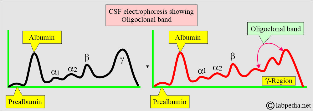
- There are several narrow bands in the gamma region of CSF electrophoresis, known as oligoclonal bands.
- The oligoclonal band is characteristic of multiple myelosclerosis.
- Ordinary cellulose acetate electrophoresis will not reveal the oligoclonal band.
- This band is seen in polyacrylamide gel, high-resolution agarose, or immunodiffusion methods.
- The oligoclonal band is seen in 85% to 90% of patients with multiple myelosclerosis.
What are the causes of oligoclonal narrow band?
- Subacute sclerosing panencephalitis.
- Destructive brain lesions.
- Brain vasculitis.
- SLE.
- Sjogren syndrome.
- Diabetes mellitus.
- Guillain-Barre syndrome.
What do you know about CSF Chloride?
- The chloride concentration in the CSF is higher than in the serum because the protein concentration in the CSF is low.
- The normal concentration is 120 to 132 meq/L.
- It falls within the CSF in cases of bacterial meningitis due to increased protein levels in the CSF.
- This test is not done routinely unless requested.
What are the causes of decreased Chloride in the CSF?
- Bacterial meningitis.
- Tuberculous meningitis.
- In a low blood chloride level.
- Its raised level is not neurologically significant; it correlates with the blood chloride level.
What do you know about CSF lactate dehydrogenase (LDH)?
- The source of LDH is neutrophils, which are responsible for fighting bacteria.
- LDH helps diagnose bacterial meningitis, particularly isoenzymes 4 and 5.
What are the causes of increased LDH in CSF?
- It is raised in CNS leukemia, with an increased cell count.
- The nerve tissue in the CNS is also high in the LDH isoenzymes 1 and 2.
- It is also raised in Stroke.
- The CSF lactate is useful in differentiating bacterial meningitis from viral meningitis.
- In viral meningitis, it is <3 mmol/L (normal range).
- >4.2 mmol/L indicates bacterial meningitis, including TB meningitis or fungal meningitis.
- This is also raised in non-Hodgkin’s lymphoma with meningeal involvement, severe cerebral malaria, head injury, and anoxia.
What do you know about CSF Lactic acid?
- CSF lactic acid does not readily pass through the blood-brain barrier; elevated blood lactic acid levels are not reflected in the CSF.
What are the causes of increased Lactic acid in CSF?
- Chronic cerebral hypoxemia or cerebral ischemia is associated with elevated CSF lactic acid levels.
- Raised in cerebral hypoxia or ischemia.
- CSF level of lactic acid increases in bacterial and fungal meningitis.
- The lactic acid level can also be increased in patients with some forms of mitochondrial diseases that affect the CNS.
- CSF level of lactic acid is normal in viral meningitis.
What do you know about CSF protein electrophoresis?
- Indication:
- Electrophoresis is used to detect any abnormalities in proteins and immunoglobulins.
- This helps to diagnose:
- Multiple sclerosis.
- Neurosyphilis.
- Autoimmune diseases.
- In Myelosclerosis, typical findings are:
- Increased total proteins, and this is mainly gamma globulins.
- The gamma region has a discrete, sharp band called the oligoclonal band.
- The oligoclonal band may be seen in HIV.
- Electrophoresis differentiates CSF from serum, as there is an additional band of transferrin in CSF that is not present in serum.
What will you see in CSF Microscopic examination?
- Normal CSF has very few mononuclear cells. Essentially free of cells.
- Normal cell count:
- Adult = 0 to 5/cmm.
- Newborn = 0 to 30/cmm.
- Child = 0 to 15/cmm.
- Neutrophils = 0 to 6% of the total cell count.
- Lymphocytes = 40% to 80 % of the total cell count.
- Monocytes = 25% to 45 % of the total cell count.
- Neutrophils in bacterial meningitis may increase from 1,000/cmm to >20,000/cmm.
What are the causes of increased Neutrophils in CSF?
- Bacterial meningitis.
- Viral meningitis.
- Tuberculous meningitis.
- Fungal meningitis.
- Amoebic encephalomyelitis.
- Abscess in an early stage.
- Metastatic tumors.
- Reaction to repeated lumbar puncture.
What are the causes of increased Lymphocytes in CSF?
- Viral meningitis.
- Syphilis with CNS involvement.
- Tuberculous meningitis.
- Multiple sclerosis.
- Guillain-barre syndrome.
- Sarcoidosis of the meninges.
- HIV.
- Fungal meningitis.
- Polyneuritis.
What are the causes of increased Monocytes in CSF?
- Chronic bacterial meningitis.
- Multiple sclerosis.
- Rupture of a brain abscess.
Can you see the Plasma cells in the CSF?
- Multiple sclerosis.
- Sarcoidosis.
- Acute viral infection.
- Infiltrated by multiple myeloma.
- Tuberculous meningitis.
- Parasitic infestation.
- Guillain-barre syndrome.
Can you see eosinophils in the CSF?
- Parasitic infestation.
- Fungal infection.
- Sarcoidosis.
- Rocky Mountain spotted fever.
Can you see the Macrophages in the CSF?
- These may be seen in TB or viral meningitis.
How will you summarize the various types of meningitis?
| Test | Bact. meningitis | TB meningitis | Viral meningitis | Fungal meningitis |
|---|---|---|---|---|
| Glucose | Decreased from 0 to 30 mg/dL | Decreased < 50 mg/dL | Normal | Normal to decreased |
| Protein | Increased ++++ | Increased ++ 50 to 300 mg/dL | Increased ++ | 50 to 300 mg/dL |
| TLC/cmm | >500 (1000 to 10,000) | Increased from 10 to 200 | increased from 10 to 200 | 40 to 400 |
| DLC | >75 % polys | >75 % lymphocytes | More lymphocytes | Lymphocytes and monocytes |
| Gram stain | Positive | Negative | Negative | Can see fungal bodies |
| Bacterial Culture | Positive | Positive for TB | Negative | Negative |
| Other tests | Pellicle or coagulum formation | Normal lactate | Indian ink |
Normal/Abnormal findings of CSF
| Lab findings | Source 1 Normal | Source 2 Normal | Abnormal findings |
|
|
||
|
<20 cm H2O |
|
|
|
|
|
|
|
|
|
|
|
|
|
|
| Differential count | |||
|
|
|
Increased =
|
|
|
|
Increased =
|
|
|
|
Increased =
|
|
Increased =
|
||
|
Increased =
|
||
| Chemistry |
|||
|
|
Increased
Decreased =
|
|
|
|
||
|
|
||
|
|
|
|
|
|
|
|
|
|
||
|
|
||
|
|
||
|
50 to 75 mg/dL (20 mg/dL less than the blood glucose level) |
60 to 80 mg/dL | Increased =
Decreased =
|
|
|
|
|
|
|
|
|
|
|
|
|
|
|
|
|
|
|
||
|
|
|
|
|
|
|
|
|
|
|
|
|
|
|
|
|
|
||
|
|
||
|
|
||
|
|
|
|
|
|
||
|
|
||
|
|
||
|
|
|
|
Questions and answers:
Question 1: What is the glucose level in bacterial meningitis?
Question 2: How to check the clarity (appearance) of the CSF?

Thanks to write and share this informative topics with very particular order .
Thanks for the encouraging remarks
why their is decrease in the Glucose in case of meningitis
If the cause is bacterial meningitis.
How to collect CSF ?
CSF collection will be done by an expert physician.
method of CSF protein estimation??
You can measure CSF protein by 1. Colorimetric method using trichloracetic acid 2. Or by the Visual semiquantitative method.
why chlroide is increased
why chlroide is decreased
CSF Chloride
The concentration of chloride in the CSF is higher than in the serum because protein concentration in the CSF is low.
The normal concentration is 120 to 132 meq/L.
It falls in the CSF in case of bacterial meningitis due to increased proteins in the CSF.
This test is not done in routine unless requested.
Decreased Chloride is seen in:
Bacterial meningitis.
Tuberculous meningitis.
In low blood chloride level.
Its raised level is not neurologically significant, it correlates with the blood chloride level.
what about potassium ?
why potassium is low in csf than serum?
you said the chloride in csf is high because protein is low so
what is the relationship between protein and chloride?
The CSF chloride concentration is higher than that of plasma because there are so negatively charged protein ions in CSF. The chloride concentration in CSF increases to balance positive charges.
The CSF chloride concentration decreases as CSF protein concentration is in pathologic states.
Very good presentation, all topics were covered and more informative.
Thanks.