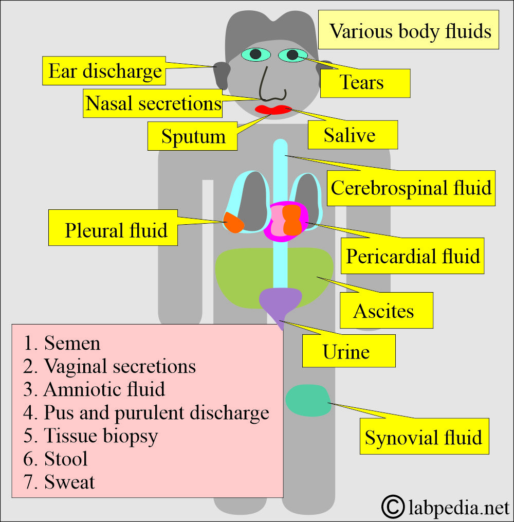
Body fluids, Their Importance, and Risk of Infections
Body fluids
How are the body fluids distributed in the body?
- The sum of fluids within all body compartments comprises total body water is about 60% of the body weight.
- It is 60% in males and 50% in females, while in infants is 70% of the total body weight.
What is the distribution of the body fluids?
- The body fluids are distributed in various compartments of the body:
- Intracellular fluid:
- It accounts for approximately 40% of the total body weight, or around 28 liters.
- Extracellular fluid:
- It is approximately 20% of the total body weight, equivalent to around 14 liters.
- This fluid may be interstitial = 15% of total body weight (11 liters).
- This may be intravascular = 5% of total body weight (3 liters).
- Total body water = 60% of the total body weight (42 liters).
- At birth, total body water is 75% to 80% of the body weight. It decreases to approximately 67% during the first year of the baby’s life.
- During adolescence, total body water accounts for approximately 60% to 65% of total body weight.
- Males have more total body water due to their increased muscle mass, while females have less due to their higher body fat content.
What is the role of Water movement between intracellular fluid (ICF) and Extracellular fluid (ECF)?
- Potassium (K+) maintains the osmotic balance of intracellular fluid (ICF).
- Sodium (Na+) maintains the osmotic balance of extracellular fluid (ECF).
What is the distribution of body fluids?
- Blood.
- Bloody fluids.
- Pleural fluid.
- Pericardial fluid.
- Peritoneal fluid (ascites).
- Amniotic fluid.
- Cerebrospinal fluid.
- Semen.
- Urine.
- Vaginal secretions.
- Saliva is a dental procedure.
- Synovial fluid.
- Pus and purulent discharge.
- Tissue biopsy or organ which is unfixed.
What are the risks and routes of infection?
- Many body fluids, including blood, are infectious and hazardous materials for humans, particularly those working in laboratories.
- These biological hazards expose unprotected laboratory workers to bacteria, viruses, parasites, and other biological agents.
- This infection may be contracted through ingestion, inhalation, inoculation, or direct contact. The infectious material may be inhaled from the patient or their body fluids or tissues.
What are the sources of contamination?
- Contaminated laboratory equipment.
- Improperly processed blood products.
- Inappropriate disposal of laboratory waste products.
What are the noninfectious materials?
- Sputum.
- Saliva.
- Nasal secretion.
- Sweat.
- Stool.
- Tears.
- Urine.
What is the importance of various body fluids?
Saliva:
- Salivary glands produce almost one liter of fluid per day, consisting of serous and mucinous material.
- Some drugs that can be monitored include Digoxin, Phenytoin, Theophylline, phenobarbital, dexamethasone, benzodiazepines, cocaine, heroin, codeine, and nicotine.
- Drug abuse, such as alcohol, barbiturates, amphetamines, and marijuana, can also be monitored.
- Hormones such as progesterone, 17-alpha-hydroxyprogesterone, cortisol, DHEA, Androstenedione, and testosterone can also be measured.
- Sjögren’s syndrome can be diagnosed by measuring increased sodium (Na+) and chloride levels.
- Radioactive material is measured for the diagnosis of Hyperthyroidism and hypothyroidism.
- Can diagnose cystic fibrosis.
- Can estimate secretory IgA.
- Take care to prevent saliva contamination from blood due to chewing or flossing.
Nasal secretions:
- It helps to diagnose allergies by showing an increased number of eosinophils.
- An increased number of polys indicates acute infection.
- Increased eosinophils and neutrophils indicate infection superimposed on chronic allergy.
- Can detect RSV by rapid antigen test.
- It will differentiate between nasal secretions and CSF in a possible skull fracture.
Sputum:
- The usefulness of the sputum culture is controversial.
- Mostly, the sputum is contaminated with bacteria from the upper respiratory tract.
- Some contaminants are Staphylococcus aureus, Streptococcus pneumoniae, Haemophilus influenzae, enteric gram-negative organisms, and Candida.
- In the literature, there is a dispute regarding whether sputum culture can identify the causative agent.
- In case a pure growth of the organism is found to favor the pathogenic infection.
- In case of increased squamous cells, favor the contamination by oropharyngeal mucosa.
- Squamous epithelial cells >19/LPF = poor specimen.
- Squamous epithelial cells 11 to 19/LPF = Fair specimen
- Squamous epithelial cells <10/LPF = Good specimen.
- The more squamous epithelial cells are found, the greater the chances for contamination.
- Sputum can be used for cytological studies to rule out malignancy.
- Sputum can diagnose various diseases:
What are the different colors of the sputum?
| Sputum character | Possible cause |
|
|
|
|
|
|
|
|
|
|
|
|
|
|
Tears:
- Tears may decrease in Sjögren syndrome, dehydration, decreased facial nerve function, and Horner syndrome.
- The deficiency of enzymes may be diagnosed in lysosomal diseases through analysis of tears.
- The following diseases are diagnosed:
- Tay-Sachs disease.
- Fucosidosis.
- Type II glycogenesis.
- Metachromatic leukodystrophy.
- Fabry disease.
- Sandhoff disease.
- Mannosidosis.
- Hurler and Scheie syndrome.
- Gm1-gangliosidosis.
- Decreased volume is also seen in dehydration.
Milk:
- If there are increased WBCs in the milk, that will indicate acute infection.
- >103 colonies of the bacteria/mL (WBC >106) indicate infections compared to the noninfectious inflammation or clogged ducts.
- It is usually due to S. aureus or S. epidermidis (penicillin-resistant).
- This infection develops in nursing mothers (∼2.5%), usually 2 to 5 weeks after postpartum.
Cerebrospinal fluid (CSF):
- CSF may be the cause of bacterial or viral meningitis.
- Infants under 1 to 2 months old are most commonly affected by infections caused by B. streptococci.
- Children from 3 to 5 or 6 years have H. Influenzae as the commonest organism, followed by meningococci and pneumococci.
- Children and older adults are more susceptible to meningococcal meningitis; the second most common organism is pneumococci.
- In older people, pneumococci are more common than meningococci.
- CNS syphilis can also be seen and diagnosed through CSF examination.
- Mycobacterial meningitis is common in children between 6 months and 5 years old. It is also seen in elderly patients.
- Cryptococcal meningitis is the most common fungus-producing CNS infection. Other fungi that cause CNS infections include Candida, blastomycosis, histoplasmosis, and coccidioidomycosis.
- HIV also produces mild aseptic meningitis.
Semen:
- There may be infection by chlamydia and syphilis.
- The presence of inflammatory cells indicates genitourinary infections.
- The most common organisms are E.coli.
- Viruses can also live in the semen, like Ebola, HIV, hepatitis B, HCV, and herpes.
- Chronic infections affect the sperm count and lead to infertility.
Vaginal secretions:
- Trichomonas causing vaginitis is quite common. The sensitivity of a freshly prepared PAP smear is 50% to 70%.
- Both aerobic and anaerobic organisms may cause bacterial infections of the vagina.
- There may be an infection from G. vaginalis and Mobilucus, accompanied by lactobacillus.
- Local causes may include endocrine disorders, poor hygiene, scabies, pinworms, foreign bodies, and local irritants.
- There may be infection by the N. gonorrhea, Chlamydia, Streptococcus group A, Staphylococcus aureus, and idiopathic infections associated with HIV.
Synovial fluid:
- Nearly 1 mL of the synovial fluid is aspirated for the culture and other studies.
- Gram stain reported 40% to 70% positive in septic arthritis.
- Gram-positive organisms are more common than gram-negative bacteria.
- Gonococcal infection is the most common cause.
- Occasionally, the synovial Rheumatoid arthritis (RA) test is positive before the serum test.
- Lactate is raised in septic arthritis and is >250 mg/dL.
- Complement is lowered in rheumatoid arthritis and SLE.
Sweat:
- This is important for the diagnosis of various diseases.
- There is a change in the electrolytes in cystic fibrosis.
- Various diseases have distinct odors, similar to those of maple syrup and certain medications.
- Sweat color is also important in various diseases, like brown in ochronosis, red in rifampin (overdose), and blue in occupational exposure to copper.
Stool:
- Stool examination is one of the noninvasive procedures to diagnose various diseases.
- The stool can be cultured to diagnose bacterial diseases, such as Salmonella, Shigella, and tuberculosis.
- A stool examination can be done to diagnose parasitic infestation.
- A stool examination can diagnose viral diseases.
Urine:
- The most common organism causing urinary tract infections (UTIs) is E. coli.
- The nitrite test is diagnostic for UTI.
- Nitrite-negative organisms are enterococci, N. gonorrhoeae, and M. tuberculosis.
What are the pathological fluids?
Ascites:
- Ascites may be seen in:
- Chronic liver disease = 81%
- Malignancies = 10%
- Congestive heart failure = 3%.
- Tuberculous ascites = 1.7%.
- Patients with dialysis = 1.0%.
- A Gram stain of the ascitic fluid reveals few bacteria in spontaneous peritonitis, but many are present due to intestinal perforation.
- This is evident in tuberculosis and certain types of gram-negative bacteria, such as Klebsiella, E. coli, and other gram-negative bacteria.
- Gram-positive bacteria, especially streptococci, may be seen.
Pleural effusion:
- The most common causes of pleural effusion are congestive heart failure and malignancies.
- Infections like tuberculosis and pneumonia are the third most common causes.
- Try to differentiate the chylous effusion, usually due to increased triglycerides.
- Pleural fluid CEA levels may increase in various malignancies and benign conditions.
Pericardial effusion:
- Pericardial effusion may develop in 40% of cases due to active rheumatic fever.
- Bacterial infections leading to pericardial effusion are caused by tuberculosis, Streptococcus pneumoniae, Staphylococcus, and gram-negative bacilli.
- Other infections may be due to Coxsackie B, rickettsial parasites, mycobacteria, and fungi.
- The most common cause is viruses.
- Rarely this may develop into severe anemia, scleroderma, polyarteritis nodosa, Wegener granulomatosis, rheumatoid arthritis, radiation, mycotic infections, and idiopathic.
Ear discharge:
- Ear discharge is also called otorrhea.
- A middle ear infection can be caused by either bacteria or viruses.
- Ear discharge may be seen in the ruptured eardrum.
- Other causes of ear discharge are swimming, foreign objects, wax, and trauma.
Questions and answers:
Question 1: What will you call ear discharge?
Question 2: What is the major cause of ascites?


