Antineutrophil cytoplasmic antibody (ANCA), Wegener’s Granulomatosis
Antineutrophil cytoplasmic antibody (ANCA)
What sample is needed for Antineutrophil cytoplasmic antibody (ANCA)?
- The patient’s serum is needed.
- How to Obtain a Good Serum Sample: Collect 3 to 5 mL of blood in a disposable syringe or a Vacutainer tube. Keep the syringe at 37 °C for 15 to 30 minutes and then centrifuge for 2 to 4 minutes to obtain a clear serum.
- No special precaution is required.
- Separate the serum as soon as possible and freeze it.
- Slides from the blood are made, and the indirect immunofluorescent technique is performed for the neutrophils.
- Two distinct staining patterns were observed for two different antibodies.
What are the Indications for Antineutrophil cytoplasmic antibody (ANCA)?
- For the diagnosis of Wegener’s granulomatosis.
- This is also done to follow the course of the disease and monitor the response to treatment of Wegener’s granulomatosis.
- This test may also be used to diagnose other autoimmune diseases.
- This test is indicated in the case of vasculitis and inflammatory bowel disease.
- This will help diagnose and classify various vasculitis-associated and autoimmune disorders.
What is the mechanism of damage by Antineutrophil cytoplasmic antibody (ANCA)?
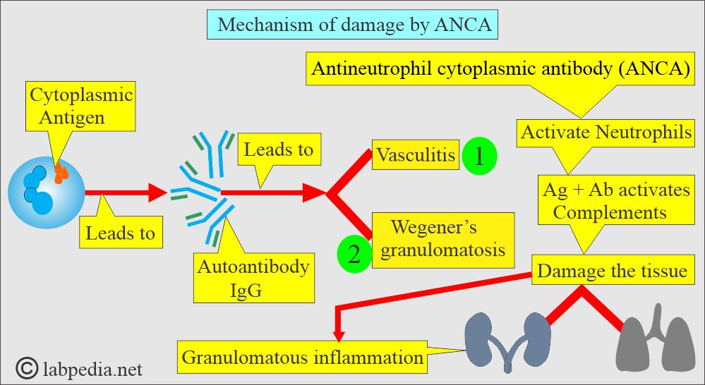
- This is also called an anti-cytoplasmic antibody. These are formed against the antigen in the cytoplasm of neutrophils and monocytes.
- The majority of these are IgG types.
- ANCA activates neutrophils and is followed by the activation of the complement pathway.
- There is damage caused by vasculitis, accompanied by a chronic inflammatory response.
- Regional vasculitis involves the small arteries of the kidneys, lungs, and upper respiratory tract.
- The pathologic damage is caused by granulomatous inflammation.
- These are autoantibodies against the myeloid-specific lysosomal enzyme.
- ANCA is seen in autoimmune diseases with vasculitis, e.g., Necrotising vasculitis, active Wegener’s granulomatosis, Polyarteritis nodosa, and renal failure.
- These are 85% to 100 % positive in Wegener’s granulomatosis.
What are the types of Antineutrophil Cytoplasmic Antibody (ANCA)?
- When patient sera are incubated with alcohol-fixed neutrophils, indirect immunofluorescence shows two patterns:
- c-ANCA(cytoplasmic ANCA):
-
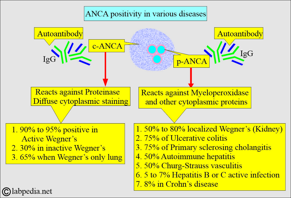
- c-ANCA (anti-proteinase 3, coarse diffuse cytoplasmic pattern) is highly specific for active Wegener’s disease.
- It is highly specific, affecting 95% to 99% of Wegener’s granulomatosis, and is characterized by diffuse granular staining of the cytoplasm of neutrophils and monocytes (using a fluorescent antibody technique).
- Its sensitivity is greater than 90% in the systemic vasculitis phase.
- It is present in only about 65% of cases of respiratory disease.
- Not all patients with limited kidney disease in Wegner’s syndrome are positive for c-ANCA (i.e., negative c-ANCA).
- When Wegner’s is not active, the percentage drops to 30%.
- A negative result for c-ANCA does not rule out Wegener’s.
- False-positive results are rare.
- A rising titer for c-ANCA suggests relapse.
- When the titer is falling, it indicates successful treatment.
- p-ANCA(perinuclear ANCA):
- It is directed against various proteins, such as myeloperoxidase, elastase, and lysozyme, and exhibits a perinuclear pattern.
- It produces a perinuclear pattern of staining in the neutrophil’s cytoplasm.
- It is effective against various proteins, including myeloperoxidase, elastase, and lysozyme.
- It is only seen in the alcohol fixation, and not in the formalin.
- ELIZA confirms a positive result,
- This test has poor specificity and a sensitivity of 20% to 60% in autoimmune diseases.
- It is found in about 50% of patients with kidney disease.
- This is also positive (approximately 75%) in other autoimmune diseases, such as ulcerative colitis or sclerosing cholangitis.
- c-ANCA appears to have anti-proteinase 3 specificity, while p-ANCA has predominantly anti-myeloperoxidase activity.
- Result: Negative when there is very little fluorescence or no fluorescence.
Wegener’s granulomatosis (Granulomatous vasculitis)
What sample is needed for Wegener’s granulomatosis?
- A biopsy of the affected tissue is taken, subjected to culture, and examined with special stains to exclude mycobacteria and fungal infections.
- Advise anti-myeloperoxidase antibody.
How will you define Wegener’s Granulomatosis?
- This is accurately referred to as granulomatosis with polyangitis.
- It is a rare autoimmune disease that causes inflammation of the blood vessels (Vasculitis), particularly affecting the sinuses, nose, kidneys, and lungs.
- It is also known as a group of disorders collectively referred to as ANCA-associated vasculitis.
- Wegener’s granulomatosis is a rare autoimmune systemic necrotizing or granulomatous vasculitis, mostly affecting the lungs and kidneys.
- Wegener’s granulomatosis is characterized by granulomatous inflammation with foci of vasculitis.
- It involves the nasopharynx, as well as a proliferative vascular lesion of the kidney.
What is the mechanism of Wegener’s granulomatosis?
- The immune system attacks small and medium-sized blood vessels.
- A proliferative vascular lesion obliterates a focal area of the Bowman’s capsule of the kidneys (crescentic glomerulonephritis).
- There is regional systemic vasculitis or fatal granulomatous vasculitis.
- Small arteries in the kidneys, lungs, and upper respiratory system (including the nasopharynx) are involved.
- The damage is due to granulomatous inflammation.
- The renal lesion is focal segmental necrotizing glomerulonephritis.
- It is a vasculitis involving small and medium-sized blood vessels in various organs.
- The common sites are the kidneys and the respiratory system.
- It may cause organ failure of the kidneys and lungs.
What are the diagnostic criteria of Wegener’s Granulomatosis?
- Necrotizing granulomas in the respiratory tract.
- Generalized necrotizing arteritis.
- Glomerulonephritis.
What are the signs and symptoms of Wegener’s Granulomatosis?
- The patient has a fever and arthralgia.
- Nosebleed, epistaxis.
- Stuffy nose, nasal ulceration, and crusty secretions.
- Rhinitis is the first symptom.
- There is sinus involvement.
- The ear shows conductive hearing loss due to auditory dysfunction.
- There is otitis media.
- The kidney shows glomerulitis.
- There is hypertension.
- The oral cavity shows nonspecific mucosal ulceration.
- There is gingivitis.
- Bone destruction and loosening of the teeth.
- The lungs show pulmonary nodules, resembling coin lesions.
- There is an infiltrate looking like pneumonia.
- There is a pulmonary hemorrhage leading to hemoptysis.
- Rarely is there bronchial stenosis.
- The skin may exhibit a subcutaneous nodule, resembling a granuloma.
- There may be cutaneous vasculitis.
- CNS may show sensory neuropathy.
- Multiple cranial nerve involvement.
- The eye shows Inflammation like:
- Scleritis and conjunctivitis are common.
- There is proptosis or ptosis.
- There are orbital granulomas.
- There is a corneal ulcer.
- There may be scleritis or episcleritis.
- There may be uveitis.
- There may be retinitis.
- Damage to the heart, lungs, and kidneys may be very severe.
- Usually, involvement of the heart, central nervous system (CNS), and gastrointestinal (GI) tract is rare.
- Histologically, there are poorly formed granulomas, characterized by the presence of necrosis and numerous multinucleated giant cells.
- It is believed that ANCA antibodies are responsible for inflammation.
How will you summarize the Clinical features of Wegener’s granulomatosis?
| Organ involved | Clinical presentations |
|
|
|
|
|
|
|
|
|
|
|
|
What are the normal ANCA levels?
- Serological method:
- Not normally found in the serum.
- Negative = <1:20
- ANCA by EIA =
- Negtive = <21 units
- Weak positive = 21 to 30 units
- Positive =>30 units.
Source 4
- Tissue biopsy
- Negative for ANCA by IFA.
- In positive c-ANCA, then the results are titrated.
- In positive p-ACNA, MPO (myeloperoxidase) testing is performed by ELISA.
- Not all p-ANCA cases are positive for MPO.
Source 2
- Blood = Negative
- ANCA is also found in ulcerative colitis and inflammatory bowel disease.
- Also positive in Crohn’s disease, specifically p-ANCA.
How will you diagnose Wegener’s Granulomatosis?
- Serological method for detecting ANCA.
- c-ANCA:
- Detection of c-ANCA is sensitive and specific for diagnosing Wegener’s disease.
- Biopsy of the affected area using the fluorescent method reveals antibody localization in the neutrophil cytoplasm, with a granular appearance throughout the cytoplasm (c-ANCA).
- c-ANCA is reported to be 90% to 95% positive in Wegner’s granulomatosis (range 84% to 100%).
- In inactive Wegner’s disease, the positivity rate is 30% (range: 13% to 41%).
- In cases involving only the respiratory system, the rate is 65% (range: 60% to 85%).
- p-ANCA reacts with myeloperoxidase and is detected in 50% to 80% of cases of localized Wegener’s granulomatosis involving the kidneys.
- p-ANCA is seen in other diseases as well, like;
- Ulcerative colitis (in approximately 75% of cases).
- Sclerosing cholangitis also shows 75% positivity.
- Autoimmune hepatitis shows 50% positivity.
- HCV and HBV in active diseases show 5% to 7% of the cases.
- Crohn’s disease shows an 8% positivity rate.
- p-ANCA is seen in other diseases as well, like;
- A biopsy is more diagnostic.
- Urine analysis reveals hematuria and proteinuria.
- Urine culture is negative.
- WBC count and ESR are raised in active disease.
- Advise chest X-Ray or CT scan.
What are the causes of increased Antineutrophil cytoplasmic antibody (ANCA)?
- Wegener’s granulomatosis (with a positive predictive value of over 90%).
- Polyarteritis nodosa
- Necrotizing vasculitis.
- Inflammatory bowel disease.
- Systemic lupus erythematosus.
- Rheumatoid arthritis.
What is the specificity of p-ANCA, and where are these seen?
- Polyarteritis nodosum.
- Primary sclerosing cholangitis.
- Microscopic polyangiitis.
- Ulcerative colitis.
Questions and answers:
Question 1: Which test is specific for Wegener's?
Question 2: What type of antibodies are found in Wegener's?


