Anemia:- Part 9 – Sideroblastic Anemia, and Anemia Due To Chronic Diseases
Sideroblastic anemia:
What sample is needed for Sideroblastic Anemia?
- EDTA blood is needed.
- Bone marrow smear for iron stain.
How will you define sideroblastic anemia?
- Sideroblastic anemia indicates a group of disorders with the presentation of:
- Anemia.
- Ineffective erythropoiesis.
- Increased serum and tissue iron.
- Increased number of ringed sideroblastic RBCs in the bone marrow aspirate (at least 20%).
- Sideroblasts are normoblasts with abnormal stainable iron in the cytoplasm that form a ring around the nucleus.
- Normoblast contains ferritin. These cells are absent in iron deficiency anemia and anemia due to chronic infection.
- Siderocytes are normal reticulocytes that contain iron in the mitochondria in the form of ferritin or hemosiderin. They are of no diagnostic importance.

Sideroblastic anemia and bone marrow show sideroblasts

Sideroblastic cells
- These anemias are due to abnormalities of heme synthesis.
How will you divide the sideroblastic anemia?
- To qualify for sideroblastic anemia, >15% of ringed sideroblasts are to be found.
- Congenital (inherited):
- This includes sex-linked congenital sideroblastic anemia.
- Autosommal recessive sideroblastic anemia.
- Acquired (drug-induced):
- Primary or idiopathic.
- Myelodysplasia.
- Secondary.
- Lead.
- Alcoholism.
- Thalassemia.
- Drugs include isoniazid and chloramphenicol.
- This may be seen in malignancies or malabsorption.
How will you discuss the pathophysiology of sideroblastic anemia?
- The pathogenesis is incompletely understood.
- This is due to abnormalities in heme metabolism.
- Several investigators found enzyme deficiency, including the deficiency of ALA synthase and uroporphyrinogen decarboxylase, in these patients.
- Irrespective of the etiology, there is an abnormal deposition of iron or siderotic granules in normoblast mitochondria. The mitochondria are present around the nucleus.
- The body has an adequate amount of iron but cannot incorporate it into hemoglobin.
- The iron (Fe) enters the developing RBCs but accumulates in the normoblast’s perinuclear mitochondria in primary sideroblastic anemia.

Sideroblast in bone marrow
How will you classify sideroblastic anemia?
A. Inherited type:
- This is rare and is sex-linked.
- Sex-linked (X-linked) anemia is more common than Autosomal recessive sideroblastic anemia.
- The gene responsible for this is ALAS2, found on the sex chromosome X.
- Pyridoxine-responsive.
- Pyridoxine-refractory.
- Autosomal, pyridoxine-refractory.
- These sideroblastic anemias are usually present in childhood.
- MCV is usually low, and RDW is usually high.
B. Acquired type:
- Primary (idiopathic).
- Myelodysplasia.
- Secondary:
- Associated with myeloproliferative disorders like leukemia and polycythemia vera.
- Pyridoxine deficiency (responsive anemia):
- Alcoholism.
- Drugs induced, such as INH, cycloserine, chloramphenicol, and chemotherapy.
- Vitamin B12 deficiency.
- Hemoglobin synthesis defects:
- Deficiency of vitamin B12 and folic acid.
- Erythropoietic porphyria.
- Lead poisoning.
- Radiation.
- Other diseases are e.g., rheumatoid arthritis, carcinoma, megaloblastic, hemolytic anemia, hemolytic anemia, and malabsorption.
- A specially acquired rare form of Pearson syndrome has sideroblastic anemia, pancreatic insufficiency, and copper deficiency.
- Pyridoxine-responsive anemia:
- Classical type.
- Variant forms.
What are the signs and symptoms of sideroblastic anemia?
- Typically, anemia appears early, usually within the first few months or years of life.
- The patients will show pallor and splenomegaly in the sideroblastic anemia.
What are the lab findings of sideroblastic anemia?
- The diagnostic feature is nucleated RBCs with iron granules called ringed sideroblasts, present in the bone marrow and dimorphic picture in the peripheral blood smear.
- Hemoglobin is low.
- MCV is variable and may be low, normal, or increased.
- MCH and MCHC are often low but may be normal.
- White blood cells may show abnormalities.
- RDW is increased.
- Serum iron is usually more than normal. There may be increased or normal iron stores.
- Ferritin level is also increased.
- The serum ferritin level is markedly raised.
- Transferrin% saturation is high.
- The serum B12 and folic acid levels are normal.
- Raised bilirubin level.
- LDH is raised.
- Decreased serum haptoglobin.
- Peripheral blood smears:
- It shows a dimorphic picture, with the presence of normocytic (normal RBCs) and microcytic and hypochromic (small-sized RBCs) RBCs. Occasional macrocytes are seen.
- Microcytosis is more common in the inherited form of sideroblastic anemia.
- Peripheral blood smears show hypochromic anemia, which is microcytic, normochromic, or macrocytic (dimorphic picture).
- The dimorphic picture is seen in the primary type. It is prominent anisopoikilocytosis.
- The microcytic form is mostly seen in the inherited form of sideroblastic anemia, while the macrocytic form is seen in the acquired form.
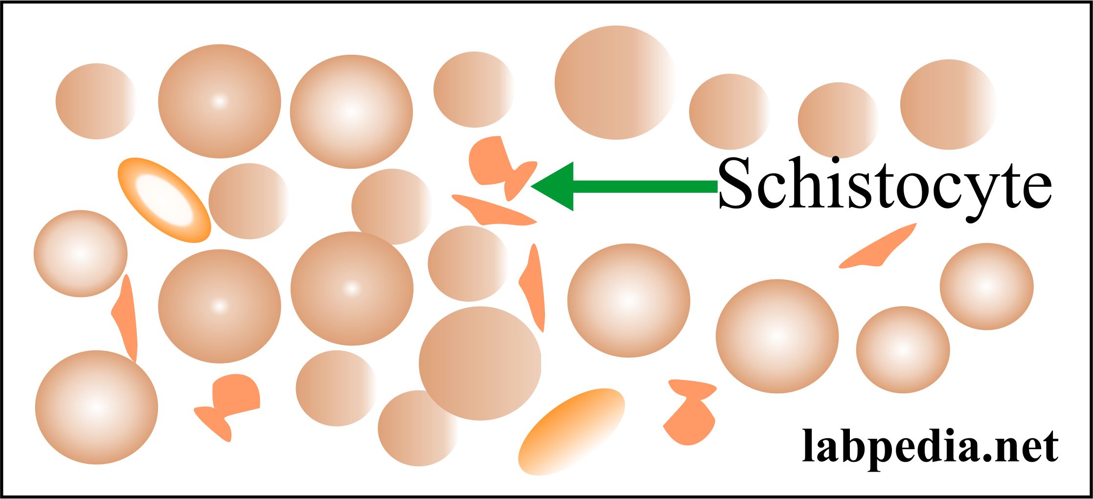
- Iron-containing Papenheimer bodies may be seen and look like basophilic stippling.

Sideroblastic anemia
- Bone marrow:
- It shows erythroid hyperplasia, but circulating reticulocytes are not increased (ineffective erythropoiesis).
- There are sideroblasts in the bone marrow aspirate. and these are >15%.
- Cytogenetics study shows a chromosomal anomaly in 25% to 50% of the cases.
- There are increased or normal iron stores.
How will you treat sideroblastic anemia?
- Treat the cause. Some patients respond when alcohol and drugs are stopped.
- In the primary type, these are unresponsive to various vitamins, especially pyridoxine (another reference says some patients respond to pyridoxine).
- Folic acid may be given in case of folate deficiency.
Anemia due to chronic diseases
How will you define anemia due to chronic diseases?
- This anemia occurs mostly in chronic inflammatory conditions and malignant diseases.
- The anemia has been present for several months following chronic diseases.
- These are commonly associated with infections, malignant neoplasms, and autoimmune disorders.
- It is usually normocytic and either normocytic or hypochromic anemia.
What is the pathogenesis of anemia due to chronic diseases?
- The mechanism of this anemia is not clearly understood.
- The basic defect is in the iron utilization for erythropoiesis.
- It looks like there is a block of iron delivery from the reticuloendothelial system to RBCs.
- A chronic disease state blocks the transfer of stored iron to maturing erythroid precursors within the bone marrow.
- This will lead to iron deficiency in RBCs while the stores have abundant iron.

Anemia due to chronic diseases
- There is a decreased release of iron from the macrophages to the plasma.
- RBCs’ life span is reduced.
- The effect of cytokines like IL-1 and tumor necrosis factor (TNF) on erythropoiesis causes an inadequate erythropoietin response to anemia.
- Hepcidin, released by the liver in response to inflammation, will inhibit macrophages’ iron release and absorption.
- The anemia will respond to successful treatment of the cause, but no response to iron therapy.
- Anemia due to chronic diseases may be due to:
- Decreased Erythropoietin response by the RBCs.
- Decreased RBC survival.
- Defective iron absorption.
- Cytokines block the release of iron from the reticuloendothelial system for the development of RBCs.
What are the causes of anemia due to chronic diseases?
- This is seen in collagen diseases (autoimmune diseases).
- Systemic lupus erythematosus.
- Rheumatoid arthritis.
- Sarcoidosis.
- Inflammatory chronic conditions.
- Tuberculosis.
- Chronic osteomyelitis.
- Fungal infection.
- Malignancies.
- Carcinoma.
- Lymphomas.
- Multiple myeloma.
What are the causes of anemia due to chronic diseases?
| Group of anemia | Causes of the chronic diseases |
|
|
|
|
|
|
|
|
What are the signs and symptoms of anemia due to chronic diseases?
- Anemia appears several months after the chronic disease.
- Anemia usually presents 1 to 3 months following the onset of chronic disease.
What are the Lab findings of anemia due to chronic diseases?
- There is Low hemoglobin (7 to 11 g/dL).
- MCV is normal.
- RBCs may be normal or microcytic and hypochromic. This microcytosis is not as severe as iron deficiency anemia.
- Usually normocytic RBCs with normal MCV, rarely MCV is <75 fl.
- In some cases, we may see hypochromic or normochromic RBCs.

Anemia of chronic diseases
- Decreased Serum iron.
- TIBC is normal or decreased.
- The serum ferritin is normal or increased.
- There is a decreased % saturation.
- Normal to increased serum ferritin level.
- It needs to differentiate from iron deficiency anemia.
- Decreased sideroblastic cells (rare to absent ringed sideroblasts).
What are the Lab findings of anemia due to chronic diseases?
| Lab test | Clinical parameters |
|
|
|
|
|
|
|
|
|
|
|
|
|
|
|
|
What are the findings in various anemias?
| Type of anemia | Hb | MCV | MCH | MCHC |
|---|---|---|---|---|
|
|
|
|
|
|
|
|
|
|
|
|
|
|
|
|
|
|
|
|
What are the characteristic findings in Various Anemias?
Anemia type |
HB |
MCV |
MCH |
MCHC |
Ferritin |
Iron binding capacity |
serum iron |
RDW |
| Iron deficiency | low | low <76 fl | low | low/normal | decreased | increased | decreased | increased |
| Megaloblastic | low | high >100 fl/cell |
increased >32 pg |
low 32 to 36 g/dL | raised/normal | increased | ||
| Chronic illness | low | low/normal | low | low | normal/ increased | normal / decreased | decreased | normal |
| Alpha Thalassemia | low or normal | low | low | low | normal /increased | normal |
normal or increased |
increased |
| Beta Thalassemia | low | low | low | low | increased/normal | normal | increased/normal | increased |
| Aplastic anemia | low | increased | normal | normal | normal |
How will you classify anemia based on RDW?
| Cell size | Normal RDW | High RDW |
|
|
|
|
|
|
|
|
|
What are the abnormalities of RBC morphology and their etiology?
| Type of RBC abnormality | Etiology of the abnormality |
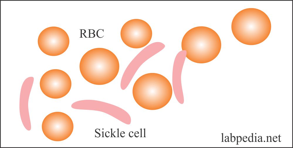
 Sickle cell Hb structure |
|
|
|
|
|
 RBC target cell |
|
|
|
|
|
|
|
|
|
|
|
|
|
|
|
|
|


How will you summarize lab findings in various anemias?
| Lab test | Iron-deficiency anemia | Pernicious anemia | Folic acid deficiency | Aplastic anemia | Thalassemia | Sideroblastic anemia | Hemolytic anemia | Post-hemorrhagic anemia | Anemia of chronic diseases |
| Hemoglobin | Low | Low | Low | Low or normal | Low | Low | Low | Normal or low | Low |
| Hematocrit | Low | Low | Low | Low or normal | Low | Low | Low | Normal or low | Low |
| MCV | Low | High | High | A normal or mild increase | Low | Low | Normal or high | Slightly low | Low or normal |
| Reticulocytes count | A normal or mild increase | Low | Low | Low | Increased | A normal or mild increase | High | Increased | Normal |
| Plasma Iron | Low | Increased | Increased | Increased | Increased or normal | Increased | Normal or high | Normal | Low |
| TIBC | Increased | Normal | Normal | Normal | Normal | Normal | Normal | Normal | Low |
| Ferritin level | Low | Increased | Increased | Normal | Increased or normal | Increased | Normal | Normal | Normal |
| Folate level | Normal | Normal | Low | Normal | Normal | Normal | Normal | Normal | Normal |
| Serum B12 level | Normal | Low | Normal | Normal | Normal | Normal | Normal | Normal | Normal |
| Transferrin | Low | Mild increase | Mild increase | Normal | Increased | Normal | Normal | Mildly low | |
| Bilirubin | Increased |
Questions and answers:
Question 1: What is the peripheral blood picture of sideroblastic anemia?
Question 2: What is the mechanism of anemia due to chronic diseases?