Androgens: Adrenal Androgens, Androstenedione (AD), DHEA-S, DHEA
Androgens
What sample is needed for Androgens?
- This may be done on 24-hour urine samples.
- Venous blood is needed to prepare the serum.
- Keep the serum on ice or cool, and perform the test within one hour.
- The serum sample can be stored at 4 °C for 2 days or months at -20 °C.
- Collect the sample from females at least one week before or after the menstrual cycle.
- Take 3 to 5 ml of blood in the disposable syringe. Keep the syringe for 15 to 30 minutes and then centrifuge for 2 to 4 minutes. This will give a clear serum.
What are the Indications for Androgens?
- This test is done to evaluate virilizing syndrome in females:
- Excessive hair growth.
- Irregular period.
- Infertility.
- This test can evaluate the adrenal glands’ function.
- This test is done to assess delayed puberty.
- D/D of Cushing syndrome (DHEA-S).
How will you define androgens?
- Androgens are sex hormones related to male characteristics present in both males and females.
- The most well-known androgen is testosterone.
- Androgens are important for sexual development.
- Androgens have a role in reproduction and overall health.
- Androgens are steroid hormones needed for the development of male characteristics.
- Androgen sources are:
- Adrenal glands are present in both males and females.
- Ovaries in females.
- Testes in the males.
How would you discuss the pathophysiology of Adrenal Androgens?
- The most important androgens are:
- Dehydroepiandrosteronene (DHEA).
- Dehydroepiandrosterone-sulfate (DHEA-SO4). It is a metabolite of DHEA.
- Daily production in young men is 30 mg/day and 20 mg/day in young women.
- Its half-life is 8 to 11 hours.
- Its half-life is 30 to 60 minutes for unconjugated androgens.
- DHEA and DHEA-S levels fall during illness, depression, and other stresses.
- Testosterone. It is the major and main hormone.
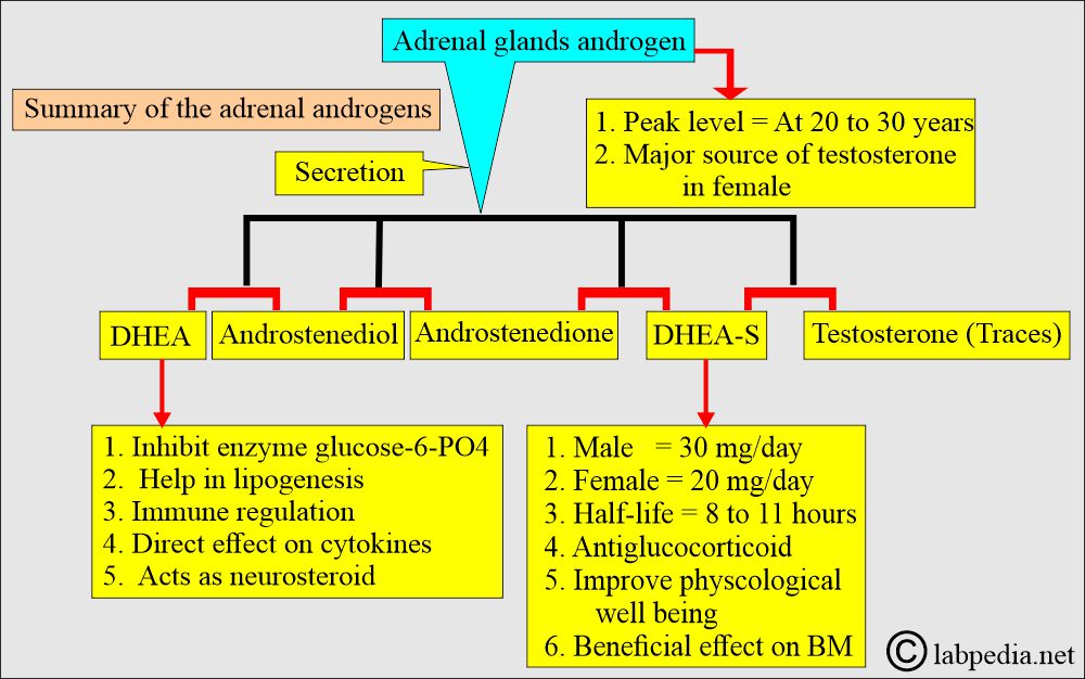
- Adrenal androgens peak between 20 and 30 years of age and then gradually fall.
- Androstenedione and DHEA are androgenic steroids produced by the adrenal cortex, ovaries, and testes.
- These are metabolically converted into testosterone and other androgens.
- Adrenal androgens are produced from the Zona fasciculta and Zona reticularis from pregnenolone and 17-OH pregnenolone.

Adrenal gland androgen hormones

Adrenal androgens and testosterone

Adrenal androgens
- The peripheral tissue converts these hormones into a relatively high level of testosterone.
- In females, androstenedione from peripheral tissues and ovaries is converted into testosterone and estrogen.

Adrenal androgen synthesis
- DHEA and DHEA-S are precursors of testosterone and estrogen produced by the gonads and adrenal glands.
- DHEA-S is produced at 8 to 16 mg/day, which is more than 90% of the plasma circulation.
- Androstenedione is elevated and gives rise to hirsutism and virilization.
Summary of Adrenal Androgens
- ACTH stimulates the secretion of the adrenal glands.

Adrenal gland regulation
What are the Adrenal androgens?
- It changes with age. It starts around 9 years of age, just before puberty onset.
- The peak level is around the third decade.
- ACTH controls the adrenal gland secretions. ACTH partially regulates the adrenal cortex secretion in adults.
- DHEA and androstenedione are secreted along with cortisol.
- Glucocorticoid therapy suppresses the secretion of adrenal androgens.
- The adrenal cortex’s average secretion is:
- DHEA = 4 mg/day.
- DHEA-S = 10 mg/day.
- Its half-life is 8 to 11 hours.
- Testosterone = 0.05 mg/day.
- Androstenedione = 1.5 mg/day.

Adrenal androgen with variation of age
What are the Androgens in females?
- The mean androgen production rate in women is:
- Testosterone = 0.25 mg/day.
- Androstenedione = 3.4 mg/day (during menstrual period).
- 1.6 mg/day during the menopausal period.
- DHT (Dihydrotestosterone) = 0.056 mg/day
- In Female testosterone:
- 50% to 60% is made from peripheral tissues.
- 30% is produced by the adrenal glands.
- 20% is produced from the ovary.
What are the Androgens in males?
- Testosterone is the main androgen in males and leads to:
- Masculinization of the male genital tract.
- Maturation of male secondary sex characteristics.
- Increase muscle bulk and bone mass.
- Increase Libido.
- Increase sexual performance in males.
- The main androgen production rate in males is:
- Testosterone = 7 mg/day.
- Androstenedione = 1.4 mg/day.
- DHT (Dihydrotestosterone) = 0.3 mg/day.

Testosterone sources in females
What are the effects of a raised level of androstenedione?
- Hirsutism.
- Change in voice
- Sterility.
- This test is done to differentiate sex character problems.
- This test may help assess delayed puberty.
- DHEA is an androgenic steroid that both men and women secrete.
How would you discuss the DHEA secretion?
- DHEA levels gradually increase during childhood and adolescence, rise rapidly after puberty, peak at age 20, and then decline.
- It decreases in the elderly more rapidly than other steroids.
- There is a moderate decrease in pregnancy.
- DHEA and Androstenedione have diurnal variations, with the highest secretion in the morning. Similar to cortisol, they secrete episodically.
- DHEA-S does not show diurnal variation and is present in the serum at much higher levels than DHEA and Androstenedione.
- Polycystic ovary (Stein-Leventhal syndrome) = High level of Androstenedione.
- Adrenal carcinoma = High level of DHEA-S.
- Cushing’s disease = Moderately raised level of DHEA-S.
- Cushing’s syndrome (a benign adrenal tumor) = Normal Androstenedione.
- Congenital Adrenal hyperplasia = Moderately raised level of DHEA-S.
How will you measure DHEA?
- DHEA is measured by gas-liquid chromatography, RIA, and gas chromatography.
What are the normal values of Androgen?
Androstenedione
- Newborn = 20 to 290 ng/dL
- Puberty = 8 to 50 ng/dL
- Male = 75 to 205 ng/dL
- Female = 85 to 275 ng/dL
- Postmenopausal = <10 ng/dL
Another source
- Premature = 80 to 446 ng/dL
- Newborn = 20 to 290 ng/dL
- 1 to 12 months = 6 to 68 ng/dL
- 10 to 17 years = 8 to 240 ng/dL
- Adult
- Male = 75 to 205 ng/dL
- Female = 85 to 275 ng/dL
- Source 2
- Male = 0.6 to 2.7 ng/mL
- 0.5 to 2.7 ng/mL
Normal Urine DHEA
- Adult male: 0.1 to 2.0 mg/day
- Adult Female: 0.1 to 1.5 mg/day
- Child: Less than 0.1 mg/day
Normal serum DHEA
Source 2
- Adult Male = 1.0 to 9.5 ng /mL
- Adult Female = 0.4 to 3.7 ng /mL
- Pregnant Female = 0.5 to 12.5 mg /mL
Another source
- Male = 180 to 1250 ng/dL
- Female = 130 to 980 ng/dL
- Urine =
- Male = <3.1 mg/24 hours
- Female = <1.5 mg/24 hours
Normal serum DHEA-S
Source 2
- Male = 280 to 640 µg /dL
- Female = 65 to 380 µg /dL
Another source
- Male = 125 to 619 µg /dL
- Female 29 to 781µg /dL
(Different literature gives different values)
What are the causes of increased levels of androgens?
- Hirsutism.
- Polycystic Ovarian Syndrome.
- Virilizing adrenal tumors.
- Precocious puberty.
- Cushing disease.
- Ectopic ACTH-producing tumor.
- Congenital adrenal hyperplasia.
What are the causes of decreased levels of androgens?
- Hyperlipidemia.
- Psychosis.
- Psoriasis.
- With the increasing age of men and women.
- Hypopituitarism.
- The patient is on glucocorticoid treatment.
Questions and answers:
Question 1: What is the main androgen in the male?
Question 2: Is there any effect of stress, depression, and illness on androgens?