Chromosome studies, Cytogenetics, Chromosome Karyotyping
Chromosome studies
What sample is needed for Chromosome studies?
- The test can be performed on almost any tissue, including:
- Blood. Leucocytes are a good source and can easily be obtained from the vein.
- Bone marrow.
- Placenta.
- Amniotic fluid (amniocentesis).
- Chorionic villous sample.
- Smears from the buccal mucosa are accessible but not as good as other sources.
- Skin.
- Tumor cells.
What are the Chromosomal karyotyping or chromosomal analysis Indications?
- This test is done to find a chromosomal defect that may lead to or is a disease risk.
- Count the number of chromosomes.
- Look for structural changes in chromosomes.
- On a couple that has a history of miscarriage or infertility. Both parents need chromosomal studies.
- In the case of two consecutive abortions.
- When there is more than one organ abnormality in the infant.
- In the case of developmental delay, intellectual disability, and growth retardation.
- To advise on ambiguous genitalia.
- In the case of mental retardation and or developmental delay, when the cause is not known.
- In the case of the family history of chromosomal translocation.
- To examine any child or baby who has unusual features or developmental delays, such as:
- Mental retardation.
- Growth retardation.
- Delayed puberty.
- Hypogonadism.
- Primary amenorrhoea.
- Ambiguous genitalia.
- Neoplasm.
- Prenatal diagnosis of congenital disease.
- Turner syndrome.
- Klinefelter syndrome.
- Down syndrome.
- In the case of spontaneous abortion, stillbirth, and neonatal death.
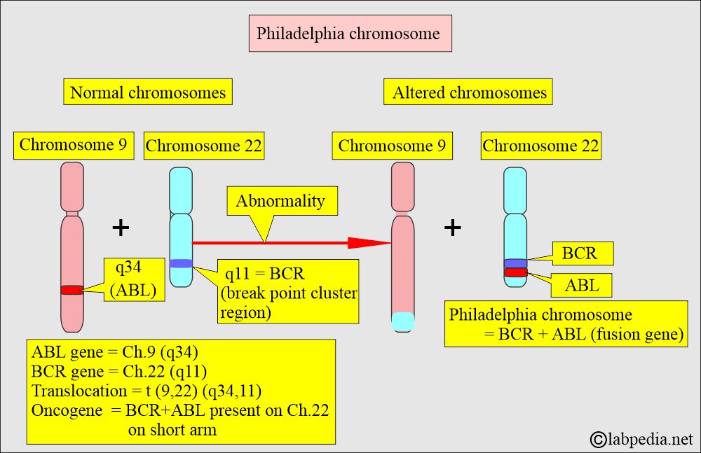
- The bone marrow or blood test can identify the Philadelphia chromosome, which is found in about 85% of people with chronic myelogenous leukemia (CML).
- The amniotic fluid test is performed to detect chromosome abnormalities in a developing baby.
- Advised women over the age of 40 years (advanced age). In these ladies, prenatal chromosomal testing is done. 1 out of 400 has a greater risk for chromosomal abnormality, biochemical abnormality, or single-gene anomaly.
- Tissue from the spontaneous abortus, stillbirth, or neonatal death is examined to find the cause of death.
What is the history of chromosomes?
- Strasburger first described chromosomes in 1815.
- Waldeyer used the term chromosome in 1888.
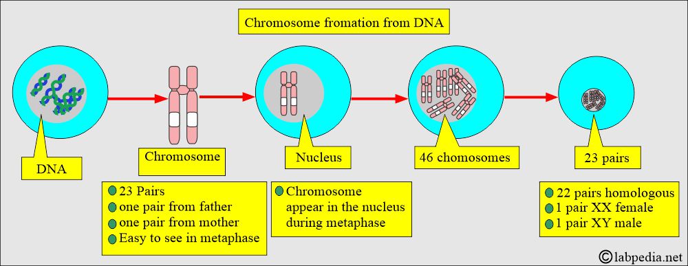
- Chromosomes appear in rod-shaped dark staining bodies during the metaphase stage of mitosis, when cells are stained with special stains and can be seen under a light microscope.
What is the effect of chromosomal abnormalities on pregnancy and the fetus?
- There may be a spontaneous abortion.
- 50% of the fertilized ova have some form of chromosomal abnormality, and >99% of these ova will die during gestation.
- There may be 50% chromosomal abnormalities in the fetus, and out of these,> 90% will die during the gestational period.
- 50% of abortions, 7% of stillbirths, and 0.5% of neonates have chromosomal abnormalities.
How is the distribution of chromosomes from both parents?
- These are diploid cells; the fetus gets one chromosome from each of the parents, one from the father, and one from the mother.
- It means the donation of one chromosome from each of the parents.
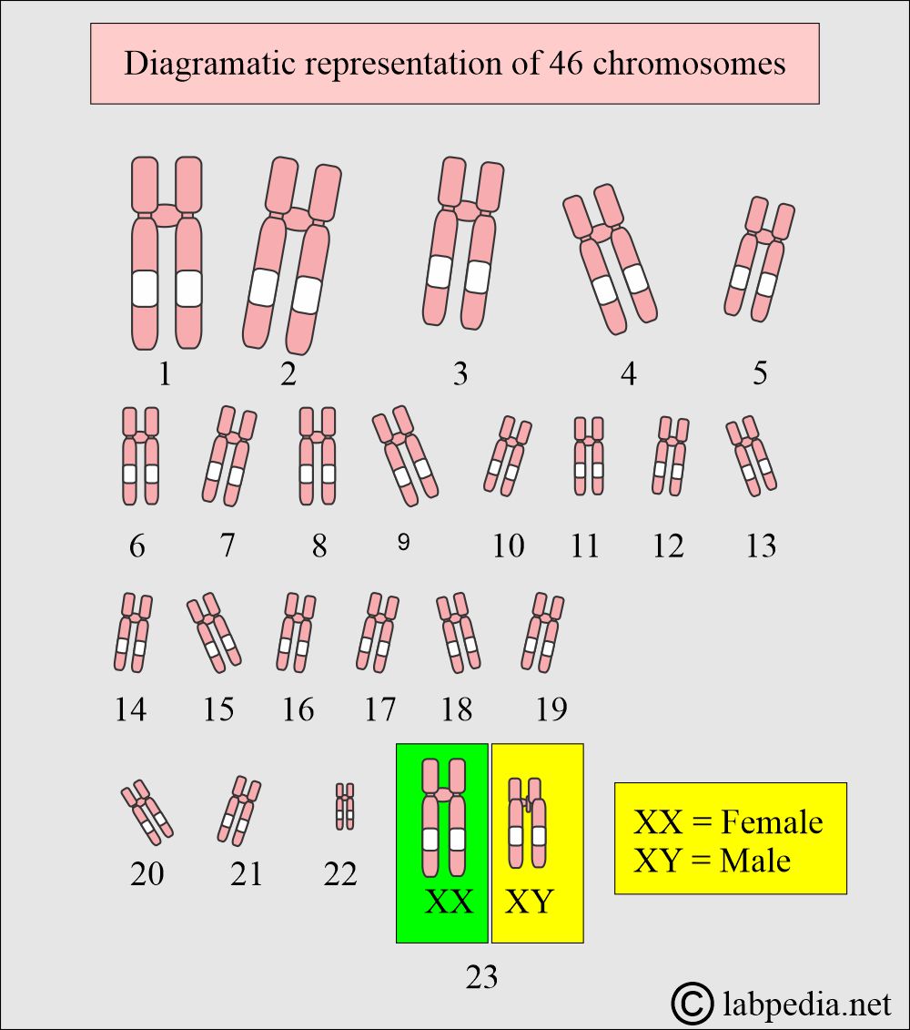
- Twenty-two chromosomes are identical, called homologous (autosomes).
- One pair consists of XX in females and XY in males.
- Chromosome number 1 is the longest.
- Chromosome number 22 is the shortest.
- Chromosomes can not be distinguished by their length because of the variation from person to person.
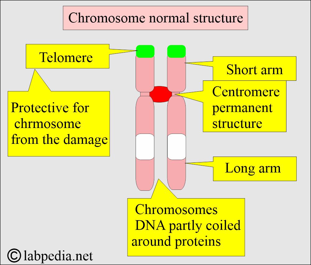
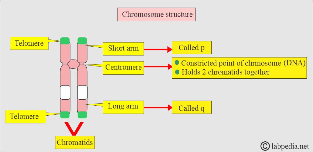
- Therefore, the position of the centromere is used to classify chromosomes.
- Chromosomes are stained with the Giemsa stain, which gives distinctive chromosome bands.
How will you describe Karyotyping?
- Karyotyping involves the separation and analysis of individual chromosomes photographed during the metaphase of mitosis.
- Karyotyping refers to the arrangement and pairing of cell chromosomes in order from the largest (longest) to the smallest (shortest) to analyze their number and structure.
- Karyotyping is the study of the number and appearance of chromosomes in the nucleus of a eukaryotic cell.
- Karyotyping refers to the arrangement of a cell’s chromosomes, their number, and structure.
- Karyotypes describe the number of chromosomes and their appearance under a light microscope.
- The normal karyotype of chromosomes consists of:
- 22 pairs of autosomal chromosomes (XX).
- One pair consists of a sex chromosome, XY for the male and XX for the female.
- The somatic cell with more or fewer than 46 chromosomes is called aneuploid.
- More than 46 chromosomes are called hyperploidy.
- A person with fewer than 46 chromosomes is called hypoploid.
What is the possibility of Chromosomal abnormalities?
- Congenital.
- Acquired.
- Chromosome abnormalities are the common cause of mental retardation and miscarriage.
- Roughly 1 in 12 conceptions has a chromosomal abnormality.
- Most of these fetuses don’t survive to term, and 50% of first-trimester fetuses abort and have major chromosomal abnormalities.
- Karyotype (Chromosomal) abnormalities occur because of:
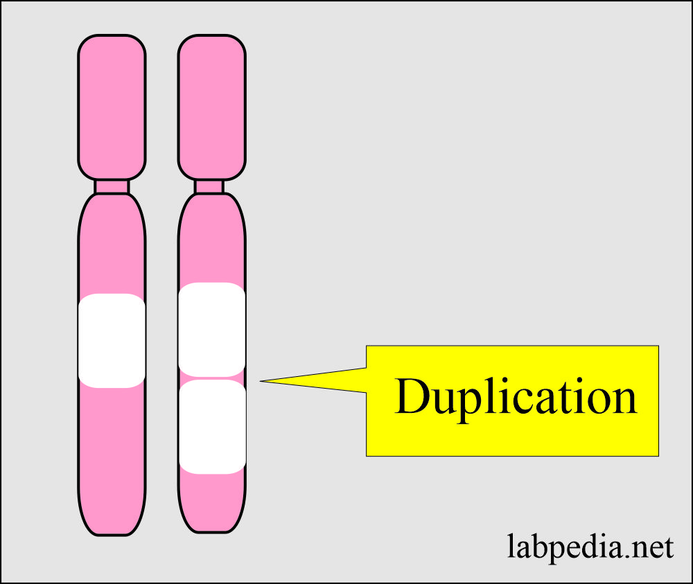
- Duplication.
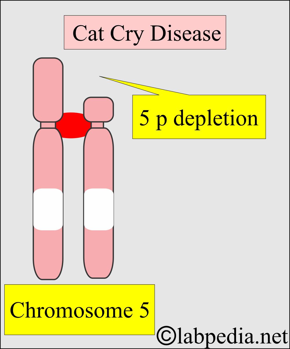
- Deletion is denoted by del. This may take place on the short or long arm.
- Translocation is denoted by t.
- Reciprocation.
- Genetic rearrangement.
What are the normal chromosomes?
- Females = 44 autosomes + 2 sex chromosomes (XX) = Karyotype = 46, XX
- Males = 44 autosomes + 2 sex chromosomes (XY) = karyotype = 46, XY
How will you perform the procedure for chromosomal analysis?
- The cells are stimulated to divide.
- Lymphocytes can be stimulated by phytohemagglutinin.
- Add Colchicine to arrest the mitotic division.
- A hypotonic solution is added to spread out the chromosomes.
- Fixation is done by Carnoy’s solution.
- Now, put the cells on the slide.
- Cell membranes are ruptured, and chromosomes remain in well-defined groups.
How will you do the Staining to see chromosomes?
- Various staining agents, such as Giemsa or Fluorescent material, stain the chromosomes.
- Giemsa banding (G banding) is the most common method for banding chromosomes.
- The dark bands are rich in DNA bases adenine and thymidine.
- The light bands are rich in cytosine and guanine.
- Quinacrine banding (Q banding) is like Giemsa banding, and it is a fluorescent method.
- Karyotyping: Now, select cells with intact chromosomes.
- Chromosomes are counted, identified, and evaluated.
- This is the arrangement and pairing of cell chromosomes, starting from the largest to the smallest in size.
- Reporting: Count autosomal chromosomes, and these are numbered according to their size.
- The larger one is labeled as 1.
How will you do the reporting of Chromosomal analysis?
- Hyperploidy is an extra chromosome.
- Hypoploidy is when the chromosomes are missing.
Duplication:
- It is when part of a chromosome is duplicated.
Deletion:
- It is when a part of a chromosome is missing.
- In addition, when the chromosome has additional material.
- Insertion is when a part of a chromosome is repositioned into a different area of the karyotype.
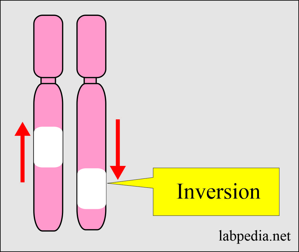
Inversion:
-
- It is when the chromosome segment is reversed in orientation.
Mosaic:
- Mosaic is when some cells have an abnormal cytogenetic abnormality, whereas the other cells don’t have it.
Translocation:
- Translocation is when there is a balanced exchange of the chromosomes without any loss or gain of material.
Chromosomal abnormalities and syndromes:
| Chromosomal abnormality | Disease or syndrome | Defect |
|---|---|---|
|
|
|
|
|
|
|
|
|
|
|
|
|
|
|
|
|
|
|
|
|
|
|
|
|
|
|
|
|
|
|
|
|
|
|
|
|
|
|
|
|
|
|
|
|
|
|
|
|
|
|
|
|
|
|
|
|
|
|
|
|
|
|
|
|
|
|
|
|
|
|
|
|
|
|
|
|
What are the possibilities of chromosomal abnormalities?
| Chromosomal abnormality | Karyotyping | Clinical presentation |
|
|
|
|
|
|
|
|
|
|
|
|
|
|
|
|
|
|
|
|
|
|
|
|
|
|
|
Questions and answers:
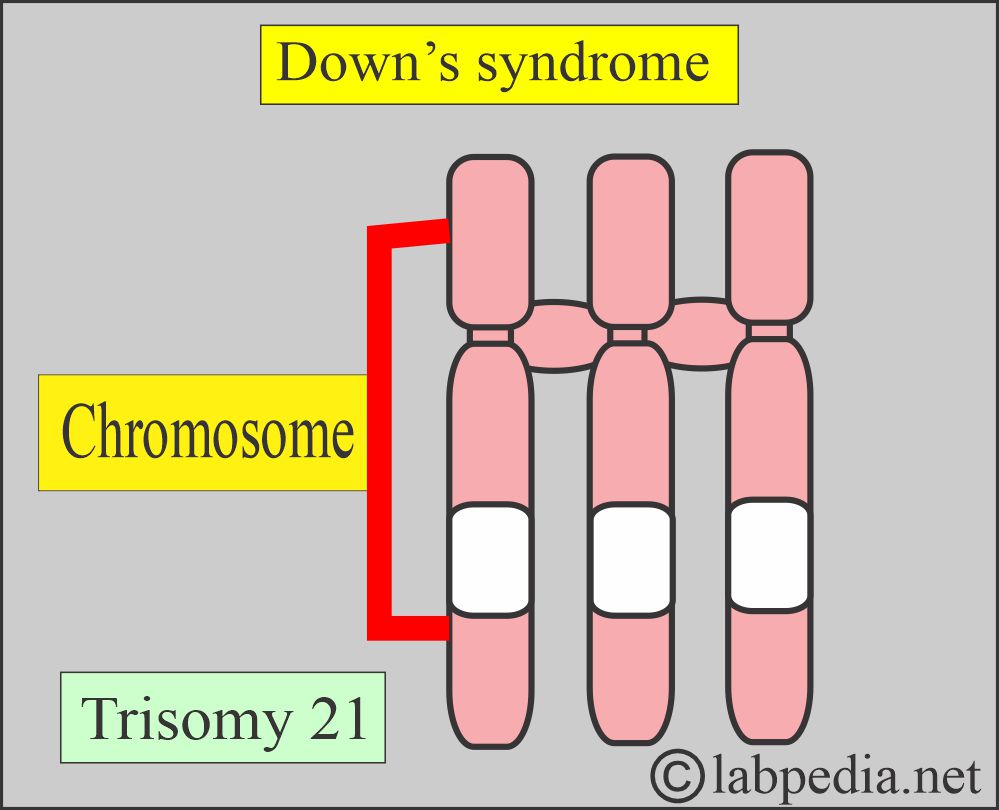
Question 1: What is the chromosomal abnormality in Down's syndrome?
Question 2: What is the chromosomal abnormality in Edward's syndrome?


Please tell me in detail all chemical reagents and stains and procedure.step by step.i think classical method will be cheaper than modren era reagents and chemical
Please clarify which stain you need step by step.
For karyotyping procedure sir