HealthFlex
×
  • Home
  • Immunology Book
  • Lab Tests
    • Hematology
    • Fluid analysis
    • CSF
    • Urine Analysis
    • Chemical pathology
    • Blood banking
    • Fungi
    • Immune system
    • Microbiology
    • Parasitology
    • Pathology
    • Tumor marker
    • Virology
    • Cytology
  • Lectures
    • Bacteriology
    • Liver
    • Lymph node
    • Mycology
    • Virology
  • Blog
    • Economics and technical
    • Fitness health
    • Mental health
    • Nutrition
    • Travel
    • Preventive health
    • Nature and photos
    • General topic
  • Medical Dictionary
  • About Us
  • Contact
March 8, 2024Lab TestsMicrobiology

Table of Contents

Toggle
  • Gram Stain
        • What sample is needed for Gram Stain?
        • What are the indications for Gram stain?
        • What is the history of Gram stain?
        • What is the structure of the bacteria?
        • How will you classify the bacteria?
      • How will you do Gram staining?
        • It consists of the following steps:
        • How will you report the results of the gram stain?
      • How will classify bacteria based on gram stain?
        • What are the examples of Bacterial Infections?
      • Questions and answers:

Gram Stain

What sample is needed for Gram Stain?

  1. Gram stain can be done on sputum, pus, tissue, and urine.
  2. The sample can be obtained from the infected ulcer or wound.
  3. The CSF may be stained.

What are the indications for Gram stain?

  1. Gram stain differentiates between gram-positive and gram-negative organisms.
  2. To diagnose the presence of bacteria in sputum, pus, or any other tissue or fluids.
  3. To diagnose bacterial meningitis.
  4. It can stain yeast, and this needs to be reported.

What is the history of Gram stain?

  1. Bacteria are colorless and usually invisible to light microscopy, so a staining color was developed to show those bacteria.
  2. The name comes from its inventor, Hans Christian Gram. He published a gram stain method in 1884.
    1. He was searching for the organism and was diagnosed with pneumonia.
  3. This is a special stain for diagnosing gram-positive or gram-negative organisms in various samples, such as sputum, pus, urine, etc.
  4. The bacteria that absorb the crystal violet and hold on to it will appear blue, called gram-positive organisms.
  5.  If the crystal violet is washed off with alcohol, it will absorb the counterstain safranin, a gram-negative organism.

What is the structure of the bacteria?

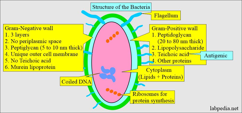
  1. Bacteria are prokaryotes (single-cell organisms).
  2. The layer outside the cytoplasm is called the peptidoglycan layer and is present in gram-positive and gram-negative bacteria.
  3.  Gram-positive bacteria have a thick wall and extensive cross-linking of the amino acid chains. In contrast, gram-negative bacteria have a very thin wall and a simple cross-linking pattern.
  4. Bacteria consist of circular DNA molecules (Continuous coding of the gene).
    1. DNA is tightly coiled.
  5. Only ribosomes are seen, which is needed for protein synthesis.
  6. Gram-positive organisms are hydrophilic, and this property prevents bacteria in the intestine from bile.
  7. Gram-negative bacteria’s outer cell walls are also hydrophilic, but the lipid component molecules give hydrophobic properties.
Bacteria structure

Bacteria structure

How will you classify the bacteria?

  1. Gram-positive = Blue.
  2. Gram-negative = Red.
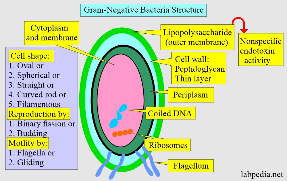
  3. The gram-negative bacterial wall consists of three layers.
Gram-Negative Bacteria

Gram-Negative Bacteria

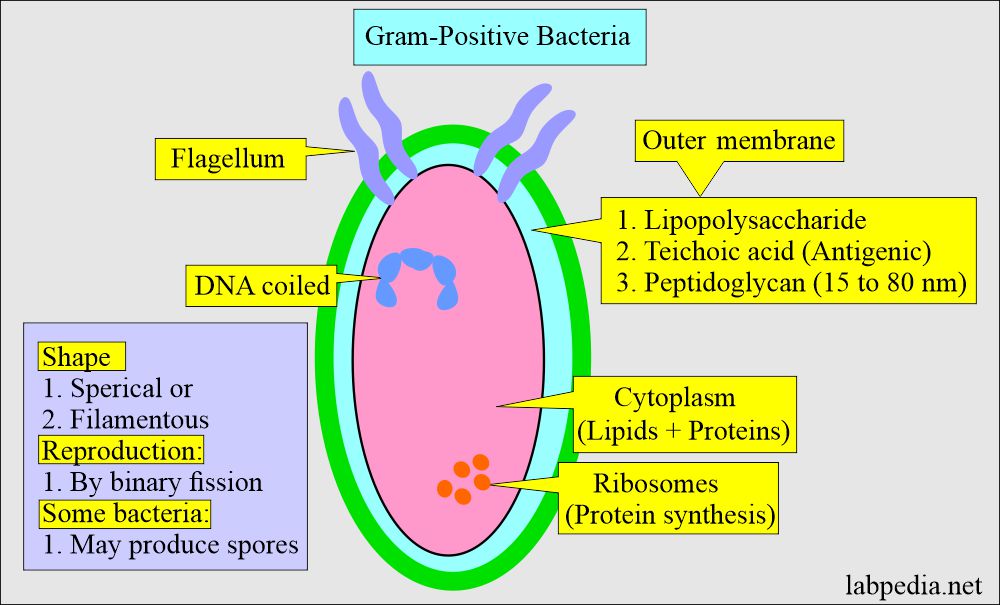
  1. Gram-positive bacteria consist of Two layers.
Gram-Positive Bacteria

Gram-Positive Bacteria

How will you differentiate Gram-positive and Gram-negative bacteria?

Characteristic features Gram-positive Gram-negative
  • Lipid contents
  • Low lipid contents
  • High lipid content
  • Number of layers
  • Two layers:
  1. Inner cytoplasmic membrane
  2. Outer thick peptidoglycan (60% to 100%)
  • Three layers:
  1. Inner cytoplasmic membrane
  2. Thin peptidoglycan (5% to 10%)
  3. Outer membrane with lipopolysaccharides
  • Teichoic acid
  • Yes
  • No
  • Peptidoglycan
  • Thick, multilayer
  • Thin, single layer
  • Periplasmic space
  • No periplasmic space
  • Positive for periplasmic space
  • Presence of toxin
  • No endotoxin
  • Endotoxin present
  • Lipopolysaccharide (Endotoxin)
  • No
  • Yes
  • Susceptibility
  • Vulnerable  to lysozyme
  • Resistant to lysozyme
  • Presence of channels (Porin)
  • No porin channel
  • There are porin channel

How will you do Gram staining?

It consists of the following steps:

  1. Fix the slide by heat.
  2. Primary stain: Stain the slide smear with gentian violet or crystal violet. It will penetrate the cell membrane.
  3. Mordant: wash the violet stain and flood the smear with the Iodine solution. This acts as a mordant and forms a complex with Crystal violet.
  4. Decolorizer: Wash off the smear and flood it with alcohol (95 %) or an Acetone-alcohol mixture.
    1. This will remove the outer cell membrane in gram-negative bacteria, where the complex will be washed off.
    2.  Gram-positive bacteria’s cell membrane remains intact, and the stain will not be washed off after alcohol treatment.
    3. Decolorization with acetone or alcohol will lead to the following:
      1. Gram-positive bacteria block the dye extraction (still, this step is unclear).
      2. This step will decolorize gram-negative bacteria and not gram-positive bacteria.
  5. Secondary stain: Counterstain the smear with safranin O, a red dye.
    1. The counterstain cannot enter gram-positive bacteria, so the bacteria are purple.
    2. In gram-negative bacteria, safranin can enter and give a pink color.
Gram stain procedure

Gram stain procedure

How will you report the results of the gram stain?

  1. Gram-positive bacteria are blue-purple.
  2. Gram-negative bacteria are pink magenta.

How will you give some examples of gram-stain bacteria?

Type of microorganism Morphology in gram stain
  • Bacillus
  • Gram + (positive) thick spore-forming bacilli
  • Streptococcus pneumoniae
  • Gram + (positive) lancet-shaped diplococci
  • Staphylococci
  • Gram + (positive) cocci as grape-like clusters
  • Streptococci
  • Gram + (positive) cocci in chain
  • Cronybacteria
  • Gram + (positive) small bacillus in Chinese letter
  • Clostridia
  • Gram + (positive) thick spore-forming spores
  • Listeria
  • Gram + (positive) small bacilli
  • Actinomyces
  • Gram + (positive) branching, filamentous, bacilli
  • Enterobacteriaceae
  • Gram – (negative) straight barrel-shaped rods
  • Vibrio
  • Gram –  (negative) curved rods
  • Neisseria
  • Gram – (negative) kidney-shaped diplococci

How will classify bacteria based on gram stain?

Bacteria  Gram-positive Gram-negative
  • Cocci
  1. Staphylococcus (Coagulase + and -)
  2. Streptococci:
    1. Group A (β-hemolyticus)
    2. Group B (β-hemolyticus)
    3. Group C (β-hemolyticus)
    4. Group F (β-hemolyticus)
    5. Streptococcus pneumoniae
    6. Streptococcus viridans
    7. Enterococcus
  1. Neisseria meningitides
  2. Neisseria gonorrhoeae
  • Bacilli
  1. Corynebacterium diphtheriae
  2. Nocardia asteroids
  3. Listeria monocytogenes
  4. Bacillus anthracis
  5. Gardnerella vaginalis
  1. Pseudomonas aeroginosa
  2. Brucella
  3. Yersinia pestis
  4. Bordetella
  5. Yersinia pestis
  6. Yesinia enterocolitica
  7. Moraxella
  8. Campylobacteria
  9. Borellia
  10. Lactobacillus

What are the examples of Bacterial Infections?

  1. Gram-positive cocci infections: 
    1. Staphylococcal aureus can cause skin infections.
    2. Toxic shock syndrome.
  2. Gram-negative cocci infections:
    1. N.meningitidis causes meningitis.
    2. N.gonorrhoeae causes gonorrhea.
  3. Gram-negative bacilli infections: 
    1. E.coli causes urinary tract infection.
  4. Gram-positive bacilli infections:
    1. B.anthracis leads to skin infection and pneumonia.
    2. Listeria monocytogenes may cause food-borne infection.
  • Viruses do not stain with gram stain.

Questions and answers:

Question 1: Who was the inventor of the gram stain?
Show answer
Hans Christian Gram invented gram stain.
Question 2: What is the role of safranin counter staining for gram-negative bacteria?
Show answer
Saranin gives pink color to gram-negative bacteria.

Possible References Used
Go Back to Lab Tests

Add Comment Cancel


The reCAPTCHA verification period has expired. Please reload the page.

  • Lab Tests
    • Blood banking
    • Chemical pathology
    • CSF
    • Cytology
    • Fluid analysis
    • Fungi
    • Hematology
    • Immune system
    • Microbiology
    • Parasitology
    • Pathology
    • Tumor marker
    • Urine Analysis
    • Virology

About Us

Labpedia.net is non-profit health information resource. All informations are useful for doctors, lab technicians, nurses, and paramedical staff. All the tests include details about the sampling, normal values, precautions, pathophysiology, and interpretation.

[email protected]

Quick Links

  • Blog
  • About Us
  • Contact
  • Disclaimer

Our Team

Professor Dr. Riaz Ahmad Bhutta

Dr. Naheed Afroz Syed

Dr. Asad Ahmad, M.D.

Dr. Shehpar Khan, M.D.

Copyright © 2014 - 2025. All Rights Reserved.
Web development by Farhan Ahmad.