Complete blood count (CBC):- Part 1 – Differential count, and Peripheral blood Smear Interpretations
Complete blood count (CBC)
What sample is needed for a Complete blood count (CBC)?
- The best sample is blood in EDTA (Ethylene diamine tetraacetic acid).
- Also, prepare fresh peripheral blood smears.
- This is inexpensive, easy to perform, and rapidly done as a screening test.
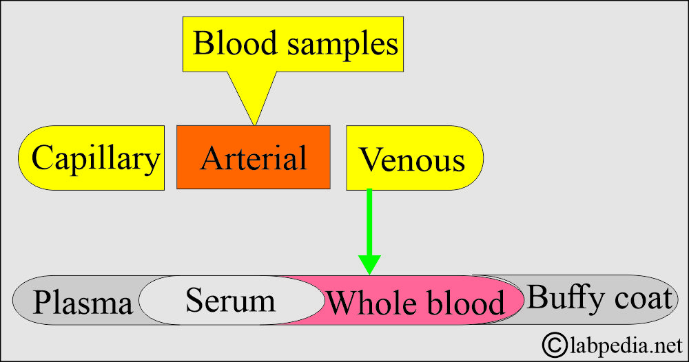
- The sample can be obtained from:
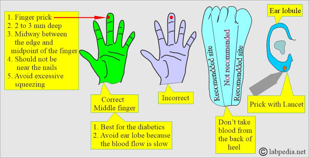
- Capillary blood can be obtained from the fingertips, heel, or big toe. This is mostly done in newborns and infants.
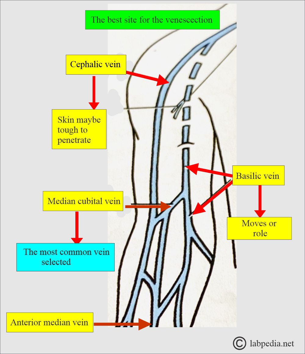
- Venous blood can be obtained from the antecubital vein, veins from the wrist area, or from any area where the vein is prominent.
What are the indications for a complete blood count (CBC)?
- It is a general screening test and gives tremendous information about the hematological system and other organ systems.
- It will differentiate between acute and chronic infection.
- It will diagnose and type the anemia.
- It will diagnose any type of leukemia.
- These are easy, inexpensive, and rapid to perform.
- It will find any abnormality in the count of platelets.
What are the precautions for a Complete blood count (CBC)?
- Physical activity and stress may cause an increase in WBCs and differential values.
- Pregnancy in the final months may cause an increase in WBC count.
- Patients with splenectomy have a persistent mild increase in the WBC count.
- Drugs that may increase the WBC count are:
- Aspirin.
- Allopurinol.
- Steroids.
- Quinine.
- Epinephrine.
- Adrenaline.
- Chloroform.
- Heparin.
- Drugs that will decrease the WBC count:
- Antibiotics.
- Anticonvulsant.
- Antimetabolites.
- Antithyroid drugs.
- Diuretics.
- Sulfonamides.
- Barbiturates.
- Chemotherapy.
How will you define Complete blood count (CBC)?
- Complete blood count (CBC) includes:
- WBC count (in 1 cmm) in the peripheral blood.
- Differential count on peripheral blood smears, with their corresponding concentration.
- RBC count and morphology.
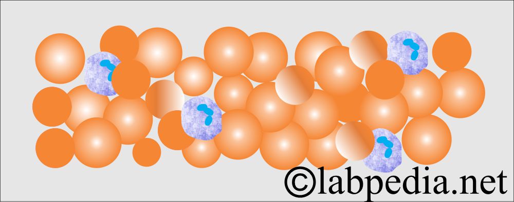
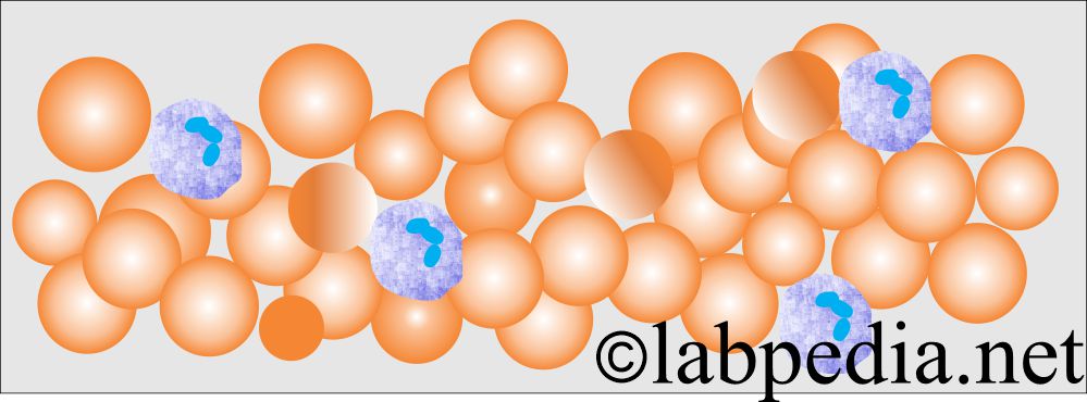
- Peripheral blood smears provide significant diagnostic information when stained with Wright and Giemsa stains.
- The best area for appreciating the different cells in the peripheral smear is one where there is no overlapping of the cells.
- Hemoglobin gives a reddish-orange appearance to the stained cells.
What are the contents of a Complete blood count (CBC)?
- This is the differential of white blood cells (polys, lymphocytes, monocytes, basophils, and eosinophils).
- Morphology of the red blood cells.
- Hemoglobin estimation (hemoglobin, which carries the oxygen).
- Hematocrit.
- Platelet count.
- Peripheral blood smears and studies include:
- RBC count.
- Hemoglobin.
- Hematocrit.
- RBC indices.
- Mean corpuscular volume (MCV).
- Mean corpuscular hemoglobin (MCH).
- Mean corpuscular hemoglobin concentration (MCHC).
- Red blood cell distribution width (RDW).
- The White cell differential count includes.
- Neutrophil count.
- Lymphocytes.
- Monocytes.
- Eosinophils.
- Basophils.
- Platelet count.
What are the advantages of a Complete blood count (CBC)?
- These are inexpensive, easy, and can be rapidly done as a screening test.
- This is the basic workup for any patient with a history of fever or any other problem to advise a complete blood count.
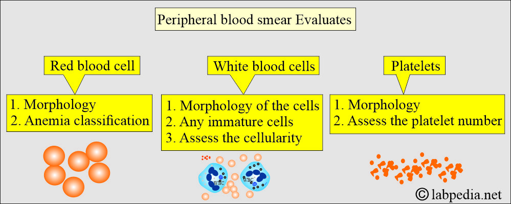
What will you see in the peripheral blood smear?
Red blood cells:
- The number of red blood cells and their morphology.
- There is minimal variation in size, shape, and staining of the RBCs, called normocytic cells.
- A variation in the size of RBCs is called anisocytosis.
- A variation in the shape of RBCs is called poikilocytosis.
- Hypochromia is a decrease in the intensity of hemoglobin staining. It is seen when the central pallor of RBCs is >1/3 of the diameter of RBCs.
What abnormalities are seen on peripheral blood smear due to RBCs?
| Type of abnormality of RBCs | Characteristics of RBCs | Importance of abnormality of RBCs |
|
|
|
|
|
|
|
|
|
|
|
|
|
|
|
|
|
|
|
|
|
|
|
|
|
|
|
|
|
|
|
|
|
|
|
|
White blood cells:
- The number of white blood cells, various types, and their morphology:
- WBCs are distinguished from RBCs by the presence of a nucleus.
- The automated machine counts all the nucleated RBCs as WBCs, so it needs to be corrected if nucleated RBCs are present in the blood.
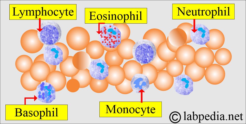
- A differential white cell count provides the proportion of different types of white cells, including neutrophils, lymphocytes, eosinophils, monocytes, and basophils.
- Visual examination of the stained slides gives the exact picture.
What is the normal differential of white blood cells?
| Type of cells | % in peripheral smear | Absolute values |
|
|
|
|
|
|
|
|
|
|
|
|
|
|
|
|
|
|
Granulocytes:
How will you define neutrophils?
- These are white blood cells that contain visible granules in their cytoplasm.
- Granules are of the following types:
- Neutrophils have neutral staining.
- Eosinophils show reddish eosinophilic granules.
- Basophils show bluish granules.
- Monocytes show a convoluted nucleus and are large in size.
What do you know about Neutrophils or polymorphonuclear leukocytes (PMN or polys)?
- Approximately 50% to 70% of the cells on the peripheral blood smear are mature granulocytes (segmented neutrophils).
- The neutrophils’ cytoplasmic granules react with both basic and acidic stains, producing neutral or light purple granules.
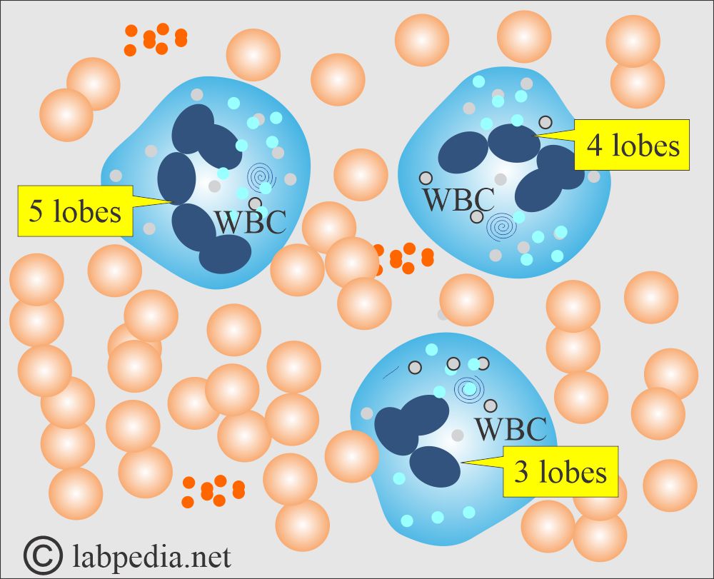
- There is a characteristic dense nucleus with 2 to 5 lobes and usually 3 lobes connected by a filament.
- The nuclear chromatin is heavily clumped, coarse, or pyknotic, and stains purplish red.
- In the mature neutrophils, the Nucleochromatin is condensed into discrete lumps or lobes.
- Roughly 6% of neutrophils have a one-lobed form called the band form.
- 35% have two lobes.
- 41% have three lobes.
- 17% have four lobes.
- 2% have five lobes.
- The cytoplasm is pale with irregular outlines containing many fine pink-blue (azurophilic) or grey-blue granules.
- The lifespan of PMNs in the peripheral blood is 6 to 10 hours.
- Band forms have less mature nuclei.
- Neutrophils are actively mobile, and many cells can gather at the site of inflammation or infection through a process called chemotaxis.
What are the functions of neutrophils?
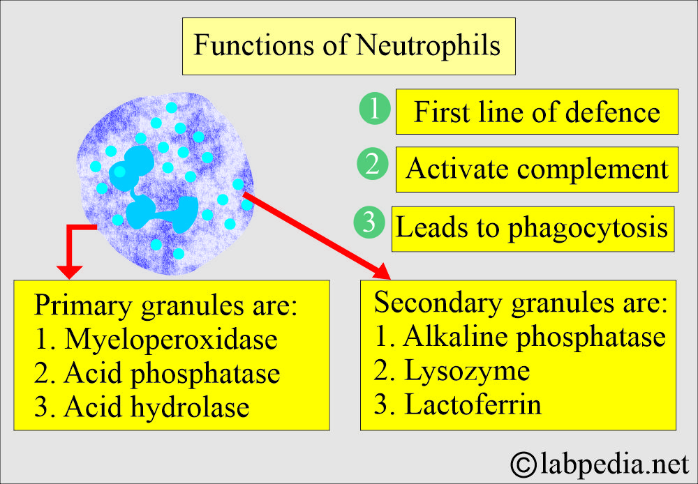
- Neutrophils are the first line of defense against tissue injury or foreign microbes.
- Segmentation of the nucleus enables these motile cells to pass through the openings of the endothelium of capillaries and target foreign substances, such as microorganisms.
- Neutrophils may activate the complement system.
- Induce immunoglobulin production.
- They can do phagocytosis and degrade some particles.
- They can produce an enzyme, which can destroy foreign substances ingested or phagocytosed.
- The neutrophil cytoplasm contains granules:
- Primary granules are myeloperoxidase, acid phosphatase, and other acid hydrolases.
- Secondary granules are alkaline phosphatase, lysozyme, and lactoferrin.
Eosinophils:
What are the facts about the eosinophils?
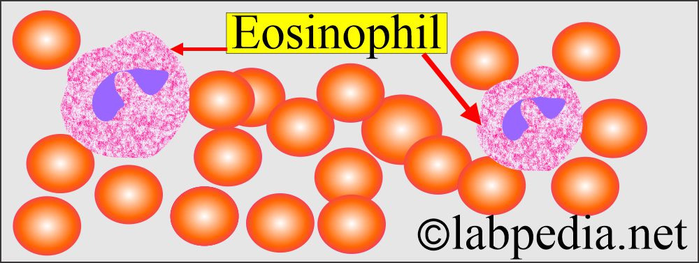
- Normal adult blood shows 0% to 4% (1% to 5%) in the peripheral blood smear.
- Eosinophils are easily recognized by eosinophilic granules in the cytoplasm and affinity for the acid eosin stain.
- In Wright’s stain, the granules are orange to reddish-orange.
- The granules are of uniform size, spherical, and evenly distributed.
- Size is slightly larger than that of the neutrophils and has two lobes with condensed chromatin. Rarely can one see three lobes.
- There is a diurnal variation in the eosinophil count, with an increase at night and a decrease in the morning.
What are the functions of eosinophils?
- These have a special role in allergic reactions.
- These are very important in parasitic defense.
- These play a role in the removal of fibrin formed during inflammation.
Monocytes:
How will you define monocytes?
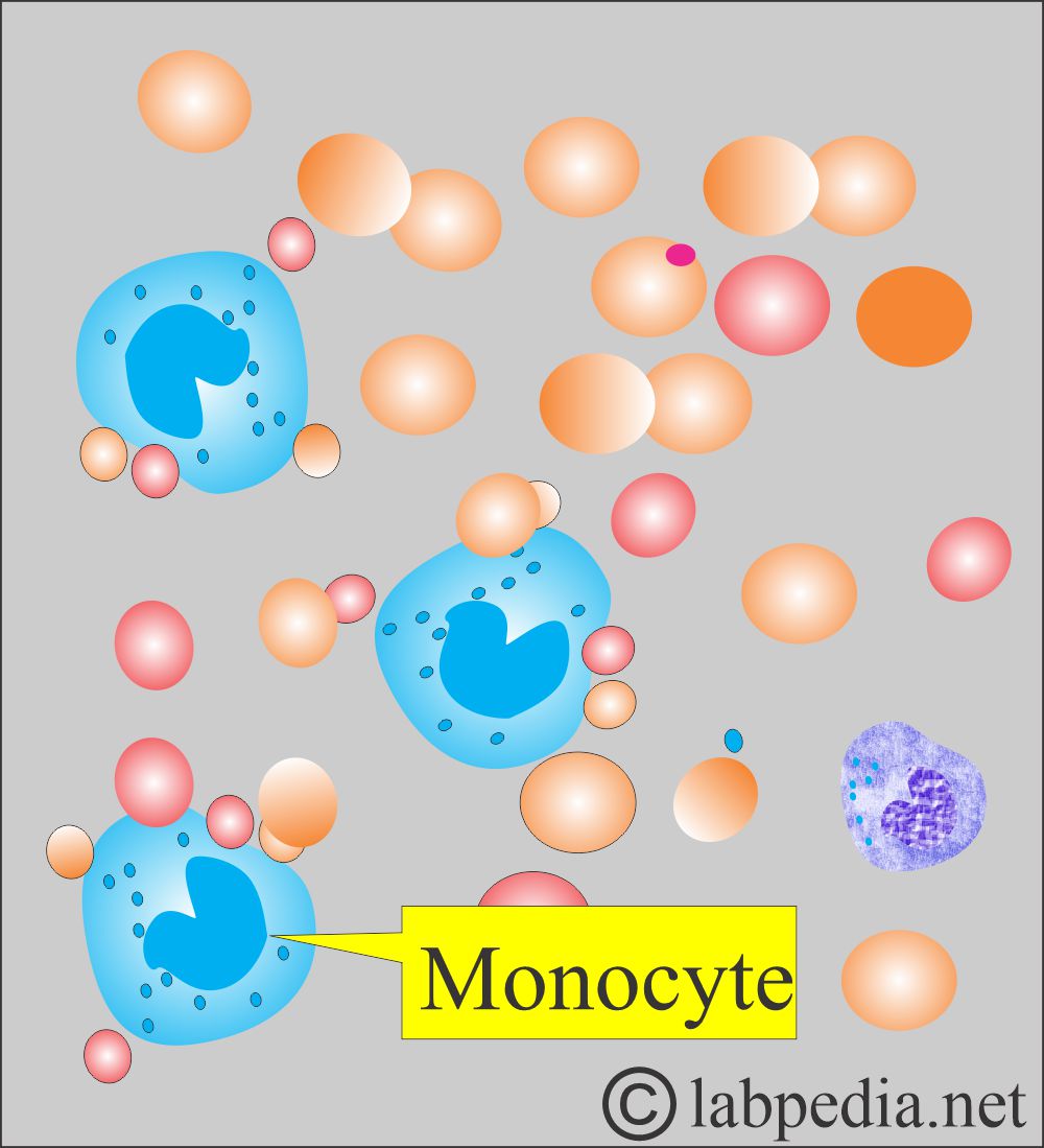
- In the DLC’s thin areas, these measure 15 to 18 µm and are larger than the neutrophils.
- There is abundant cytoplasm compared to the nucleus ( N: C = 2:1).
- With Wright’s stain, the cytoplasm turns dull grey-blue compared to the pink cytoplasm of the neutrophils.
- The nuclei of monocytes may be kidney-shaped, folded, indented, or occasionally lobulated.
- The nucleus exhibits convolutions similar to those found in the brain or a turban.
- The shape of monocytes is variable, ranging from round to those with blunt pseudopodia.
- Four important features of the monocytes are:
- Nuclear convolutions.
- Lacy or often delicate chromatin.
- Dull grey-blue cytoplasm.
- Blunt pseudopods.
- Digestive vacuoles may be seen in the cytoplasm.
- The half-life ranges from 8 hours to 3 days before these cells enter the tissues and differentiate into macrophages.
- Monocytes are 2% to 9% of the normal blood leucocytes.
What are the functions of monocytes?
- Defensive mechanism:
- Under various conditions, such as infection by bacteria, fungi, pigments, and phagocytosed RBCs, may be observed in the cytoplasm.
- Phagocytosis.
Basophils:
How will you discuss basophils?
- These cells are rarely found in the peripheral smear, accounting for 0 to 2% of the total.
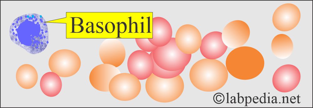
- Basophils have large, abundant violet-blue or purple-black granules in the cytoplasm.
- These granules mostly obscure the nucleus.
- These granules measure 0.2 to 1.0 µm. These vary in number, size, and shape—these are less numerous than the eosinophil’s granules.
- These granules have an affinity for the blue or basic thiazine dyes. Basophil granules are water-soluble.
- Basophils exhibit diurnal variation, similar to eosinophils, with an increase at night and a decrease in the morning.
What are the functions of basophils?
- In the tissue, they become mast cells.
- These cells have a receptor for IgE.
- On degranulation, it produces histamine.
Lymphocytes:
How will you discuss the lymphocytes?
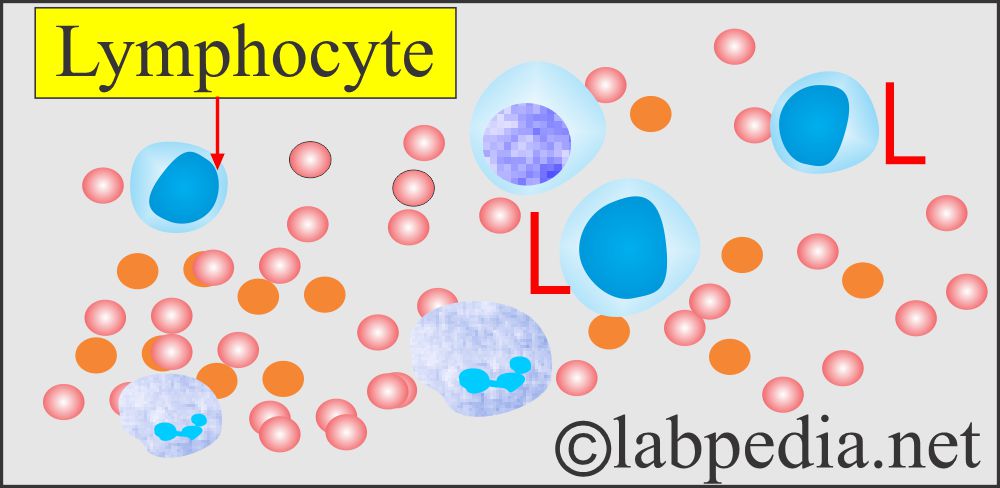
- Lymphocytes are the second most numerous cells in the peripheral blood smears.
- Lymphocytes are 20% to 40% of adult blood cells.
- Lymphocytes are small, varying in size from 7 to 10 µm. There are intermediate and large lymphocytes.
- Wright’s stain yields a blue cytoplasm, varying in intensity from light to dark, in different cells.
- Most lymphocytes don’t have granules.
- The nucleus size in small lymphocytes is just the size of RBCs in the same microscopic field.
- The nucleus /cytoplasm ratio is 4:1, and nuclei are round or slightly indented.
- Chromatin structure is lumpy or clumped and stains dark purple with a lighter bluish-purple area between chromatin aggregates.
What are the functions of lymphocytes?
- Their primary function is humoral immunity.
- B-L produces an antibody-dependent immune reaction.
- T-lymphocytes help B-lymphocytes and give rise to cell-mediated immunity.
Platelets:
How will you discuss the platelets?
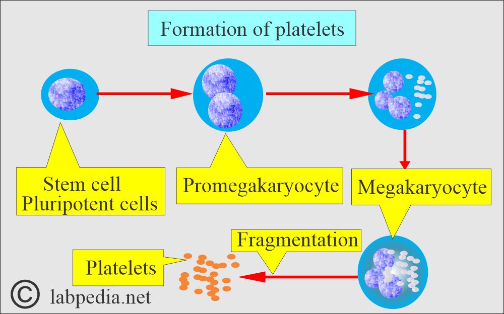
- Platelets are produced in the bone marrow by the fragmentation of the cytoplasm of megakaryocytes.
- Each megakaryocyte gives rise to 1000 to 5000 platelets.
- The size of platelets ranges from 2 to 4 µm in diameter and varies in shape.
- The average number of platelets is 7 to 15/oil immersion field.
- Estimate the number of platelets in 10 oil immersion fields.
- Platelets are mostly seen in groups.
- It is necessary to count the number of platelets in the group.
- Platelets have no nucleus.
- Normal platelet count is 250 x 10^9/L, ranging from 150 to 400 x 10^9/L.
- The normal lifespan of platelets is 7 to 10 days. About 1/3 of the marrow output of platelets is trapped in the spleen, and it will increase in the case of splenomegaly.
What is the structure of the platelets?
- The platelets are the smallest structures in the peripheral blood smear, measuring 3.0 x 4.0 µm in diameter with a mean volume of 7 to 10 fl.
- The larger platelets are greater than 4.0 µm.
- Giant platelets measure >7.0 µm.
What are the functions of platelets?
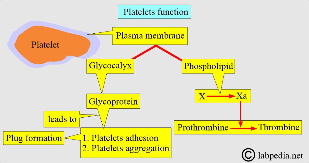
- The surface coat’s glycoproteins are crucial in the platelet’s reactions of adhesion and aggregation, which are the initial events in platelet plug formation during the process of hemostasis.
- The plasma membrane phospholipids (known as platelet factor 3) are important in the conversion of coagulation factor X to Xa and prothrombin (factor II) to thrombin (factor IIa).
- The platelets contain three types of granules:
- α-granules:
- Contains a heparin antagonist.
- Platelet-derived growth factor (PDGF).
- β-thromboglobulin.
- Fibrinogen.
- VWF and other factors.
- Dense granules contain:
- Adenosine diphosphate (ADP).
- Adenosine triphosphate (ATP).
- 5-hydroxytryptamine (5-HT).
- Calcium.
- Lysosomes contain:
- Hydrolytic enzyme.
- Peroxisomes contain catalase.
What is the Cytochemistry of various cells found in the peripheral blood?
| Types of cells | Peroxidase | Nonspecific esterase | Periodic Acid-Schiff (PAS) | Acid phosphatase |
| Segmented neutrophils | Positive (++) | Positive (+) | Positive (++) | Diffusely positive |
| Band neutrophils | Positive (++) | Positive (+) | Positive (+ to ++) | Diffusely positive |
| Eosinophils | Positive (++) | Negative | Negative (Cytoplasm +) | Diffusely positive |
| Basophils | Partly positive | Negative | Positive (++) | Negative |
| Monocytes | Negative to positive | Positive (++) | Diffusely positive (+) | Positive (++) |
| Lymphocytes | Negative |
Focal locally positive (+) (Acid esterase ) |
Granules partly positive (+) | Often positive (+), Fine granules, and focal |
| Platelets | Negative | Positive (+) | Positive (+) | Positive (+) |
What is the normal value of the cells in a Peripheral blood smear?
Normal Peripheral blood (CBC):
- TLC is the total leucocyte count:
- 4,100 to 10900/cmm.
- DLC is a differential count. Normal values are:
- Neutrophils = 48% to 77%
- Lymphocytes = 10% to 40%
- eosinophils = 0.3% to 7%
- Monocytes = 0.6% to 9.6%
- Basophils = 0.3% to 1%
- Platelets = 140,000 to 400,000 /cmm
What is the normal Hemoglobin?
Source 2
- Male 14 to 18 g /100 ml
- Female 12 to 16 g/ 100 ml
- Pregnant female = >11 g/dL
- Old people’s values are slightly low.
Source 1
| Age | Hb g/dL | Hb g/dL Male | Hb g/dL Female |
| Fetal blood | |||
| 18 to 20 weeks | 11.47 ± 0.78 | ||
| 21 to 22 weeks | 12.28 ± 0.89 | ||
| 23 to 25 weeks | 12.40 ± 0.77 | ||
| 26 to 30 weeks | 13.35 ± 1.17 | ||
| Infants | |||
| Cord blood | 13.5 to 20.5 | ||
| 1 month | 10.7 to 17.1 | ||
| 2 month | 9.0 to 13.0 | ||
| 4 month | 10.3 to 14.1 | ||
| 9 month | 11.4 to 14.0 | ||
| one year | 11.3 to 14.1 | ||
| 2 to 5 years | 11.0 to 14.0 | ||
| 5 to 9 years | 11.5 to 14.5 | ||
| 9 to 12 years | 12.0 to 15.0 | Male | Female |
| 12 to 14 years | 12.0 to 16.0 | 11.5 to 15.0 | |
| 15 to 17 years | 11.7 to 16.6 | 11.7 to 15.3 | |
| 18 to 44 years | 13.2 to 17.2 | 11.7 to 15.5 | |
| 45 to 64 years | 13.1 to 17.2 | 11.7 to 16.0 | |
| 65 to 74 years | 12.6 to 17.4 | 11.7 to 16.1 |
- For conversion into SI units x 10 = g/L
What is the critical value? = <5 g/dL or >20 g/dL
What is the normal ESR (erythrocyte sedimentation rate)?
Normal
- Male Older than 50 years = 0 to 15 mm/ hour
- Female = 0 to 25 mm /hour
- Child = up to 10 mm/hour
- Newborn = 0 to 2 mm/hour
What is the normal Platelet count?
- Adult = 140,000 to 400,000/cmm
- Children = 150,000 to 450,000/cmm
- Platelets are assessed by either a smear or a hematology analyzer.
What are the causes for increased Neutrophils?
- Infections.
- Myocardial infarction.
- Stress.
- Metabolic diseases.
- Inflammations.
What are the causes of decreased Neutrophils?
- In radiation therapy or chemotherapy.
- Infections.
- Hypersplenism.
- Folic acid or B12 deficiency.
- Hepatic diseases.
- Drugs
- collagen vascular diseases.
What are the causes of increased Eosinophils?
- Allergy.
- Parasitic infestation.
- Skin disorders.
- Neoplastic diseases like Hodgkin’s lymphoma.
- Collagen vascular diseases.
What are the causes of decreased eosinophils?
- Cushing’s syndrome.
- Stress.
What are the causes of increased Lymphocytes?
- Chronic infections.
- Lymphocytic leukemia.
- In immune diseases, e.g, Ulcerative colitis.
What are the causes of decreased Lymphocytes?
- Chronic debilitating illness.
- Immune-deficiency.
What are the causes of increased Monocytes?
- Infections.
- Collagen vascular diseases.
- Carcinoma.
- Monocytic leukemia.
- Lymphoma.
What are the causes of increased Basophils?
- Chronic myelocytic leukemia.
- Polycythemia vera.
- Hodgkin’s disease.
- In some anemias.
What are the causes of decreased Basophils?
- Hyperthyroidism.
- Stress.
- Ovulation.
- Note: For more information, please refer to the peripheral blood smear.
Questions and answers:
Question 1: What is the effect of hyperthyroidism on basophils?
Question 2: What are the causes of decreased eosinophil count?

Good subject
Recently reported in a study in JAMA that megakarocytes have been found in capillaries of brain on autopsy in Covid19 patients. How they reach these capillaries?I simply don’t understand
This is really confusing for the researcher. Please read this reference.
https://www.medpagetoday.com/neurology/generalneurology/91246
Plz send
May I know what you want?
what does slightly increased platelet distribution mean? peripheral blood smear shows: poikilocytosis, sickle cells and occasional target cells, hypochromic and microcytic cells, and normal WBC morphology.
Please see this comment:
Platelet distribution width (PDW) is a measurement of platelet anisocytosis calculated from the distribution of individual platelet volumes. Thrombocrit (or plateletcrit) is the percentage of blood volume occupied by platelets and is an assessment of circulating platelet mass.